 Movie
Movie Controller


+ Open data

- Basic information
| Entry |  | |||||||||
|---|---|---|---|---|---|---|---|---|---|---|
| Title | Structure of human NHE6.1 bound to PIP2 | |||||||||
 Map data Map data | cryoSPARC final non-uniform refinement map blurred with a bfactor of 40 for refinement of full-length model | |||||||||
 Sample Sample |
| |||||||||
 Keywords Keywords | Na/H exchanger / sodium transport / proton transport / endosome / MEMBRANE PROTEIN | |||||||||
| Function / homology |  Function and homology information Function and homology informationDefective SLC9A6 causes X-linked, syndromic mental retardation,, Christianson type (MRXSCH) / Sodium/Proton exchangers / dendrite extension / potassium:proton antiporter activity / axon extension / sodium:proton antiporter activity / sodium ion import across plasma membrane / establishment of cell polarity / neuron projection morphogenesis / potassium ion transmembrane transport ...Defective SLC9A6 causes X-linked, syndromic mental retardation,, Christianson type (MRXSCH) / Sodium/Proton exchangers / dendrite extension / potassium:proton antiporter activity / axon extension / sodium:proton antiporter activity / sodium ion import across plasma membrane / establishment of cell polarity / neuron projection morphogenesis / potassium ion transmembrane transport / regulation of intracellular pH / sodium ion transmembrane transport / recycling endosome / recycling endosome membrane / late endosome membrane / early endosome membrane / endoplasmic reticulum membrane / identical protein binding / plasma membrane Similarity search - Function | |||||||||
| Biological species |  Homo sapiens (human) Homo sapiens (human) | |||||||||
| Method | single particle reconstruction / cryo EM / Resolution: 2.55 Å | |||||||||
 Authors Authors | Hansen JS / Pike ACW / Chi G / Wolf G / Ingles-Prieto A / Tranberg-Jensen J / Ye M / Speedman D / Goericke F / Sauer DB ...Hansen JS / Pike ACW / Chi G / Wolf G / Ingles-Prieto A / Tranberg-Jensen J / Ye M / Speedman D / Goericke F / Sauer DB / Beck H / Superti-Furga G / Huber KVM | |||||||||
| Funding support |  Switzerland, 2 items Switzerland, 2 items
| |||||||||
 Citation Citation | Journal: To Be Published Title: Structure of human NHE6.1 bound to PIP2 Authors: Hansen JS / Pike ACW / Chi G / Wolf G / Ingles-Prieto A / Tranberg-Jensen J / Ye M / Speedman D / Goericke F / Sauer DB / Beck H / Superti-Furga G / Huber KVM | |||||||||
| History |
|
- Structure visualization
| Supplemental images |
|---|

- Downloads & links
-EMDB archive
| Map data | emd_53831.map.gz | 166.9 MB | EMDB map data format | |
|---|---|---|---|---|
| Header (meta data) | emd-53831-v30.xmlemd-53831.xml | 25.2 KB 25.2 KB | Display Display | EMDB header |
| FSC (resolution estimation) | emd_53831_fsc.xml | 11.8 KB | Display | FSC data file |
| Images |  emd_53831.png emd_53831.png | 112.6 KB | ||
| Masks | emd_53831_msk_1.mapemd_53831_msk_2.map | 178 MB 178 MB | Mask map | |
| Filedesc metadata | emd-53831.cif.gz | 7.7 KB | ||
| Others | emd_53831_additional_1.map.gzemd_53831_half_map_1.map.gzemd_53831_half_map_2.map.gz | 88.8 MB 165.3 MB 165.3 MB | ||
| Archive directory |  https://data.pdbj.org/pub/emdb/structures/EMD-53831ftp://data.pdbj.org/pub/emdb/structures/EMD-53831 https://data.pdbj.org/pub/emdb/structures/EMD-53831ftp://data.pdbj.org/pub/emdb/structures/EMD-53831 | HTTPS FTP |
-Related structure data
| Related structure data |  9r8mMC M: atomic model generated by this map C: citing same article ( |
|---|---|
| Similar structure data |
-Links
| EMDB pages | EMDB (EBI/PDBe) / EMDataResource |
|---|
-Map
| File | Download / File: emd_53831.map.gz / Format: CCP4 / Size: 178 MB / Type: IMAGE STORED AS FLOATING POINT NUMBER (4 BYTES) | ||||||||||||||||||||||||||||||||||||
|---|---|---|---|---|---|---|---|---|---|---|---|---|---|---|---|---|---|---|---|---|---|---|---|---|---|---|---|---|---|---|---|---|---|---|---|---|---|
| Annotation | cryoSPARC final non-uniform refinement map blurred with a bfactor of 40 for refinement of full-length model | ||||||||||||||||||||||||||||||||||||
| Projections & slices | Image control
Images are generated by Spider. | ||||||||||||||||||||||||||||||||||||
| Voxel size | X=Y=Z: 0.932 Å | ||||||||||||||||||||||||||||||||||||
| Density |
| ||||||||||||||||||||||||||||||||||||
| Symmetry | Space group: 1 | ||||||||||||||||||||||||||||||||||||
| Details | EMDB XML:
|
 Z (Sec.)
 Y (Row.)
 X (Col.)


-Supplemental data
-Mask #1
| File | emd_53831_msk_1.map | ||||||||||||
|---|---|---|---|---|---|---|---|---|---|---|---|---|---|
| Projections & Slices |
| ||||||||||||
| Density Histograms |


-Mask #2
| File | emd_53831_msk_2.map | ||||||||||||
|---|---|---|---|---|---|---|---|---|---|---|---|---|---|
| Projections & Slices |
| ||||||||||||
| Density Histograms |


-Additional map: cryoSPARC final unsharpened non-uniform refinement map
| File | emd_53831_additional_1.map | ||||||||||||
|---|---|---|---|---|---|---|---|---|---|---|---|---|---|
| Annotation | cryoSPARC final unsharpened non-uniform refinement map | ||||||||||||
| Projections & Slices |
| ||||||||||||
| Density Histograms |


-Half map: cryoSPARC final non-uniform refinement halfmap1
| File | emd_53831_half_map_1.map | ||||||||||||
|---|---|---|---|---|---|---|---|---|---|---|---|---|---|
| Annotation | cryoSPARC final non-uniform refinement halfmap1 | ||||||||||||
| Projections & Slices |
| ||||||||||||
| Density Histograms |


-Half map: cryoSPARC final non-uniform refinement halfmap2
| File | emd_53831_half_map_2.map | ||||||||||||
|---|---|---|---|---|---|---|---|---|---|---|---|---|---|
| Annotation | cryoSPARC final non-uniform refinement halfmap2 | ||||||||||||
| Projections & Slices |
| ||||||||||||
| Density Histograms |


- Sample components
-Entire : NHE6.1 homodimer
| Entire | Name: NHE6.1 homodimer |
|---|---|
| Components |
|
-Supramolecule #1: NHE6.1 homodimer
| Supramolecule | Name: NHE6.1 homodimer / type: complex / ID: 1 / Parent: 0 / Macromolecule list: #1 |
|---|---|
| Source (natural) | Organism: Homo sapiens (human) |
| Molecular weight | Theoretical: 170.856 KDa |
-Macromolecule #1: Isoform 2 of Sodium/hydrogen exchanger 6
| Macromolecule | Name: Isoform 2 of Sodium/hydrogen exchanger 6 / type: protein_or_peptide / ID: 1 / Number of copies: 2 / Enantiomer: LEVO |
|---|---|
| Source (natural) | Organism: Homo sapiens (human) |
| Molecular weight | Theoretical: 85.507812 KDa |
| Recombinant expression | Organism: Homo sapiens (human) |
| Sequence | String: MARRGWRRAP LRRGVGSSPR ARRLMRPLWL LLAVGVFDWA GASDGGGGEA RAMDEEIVSE KQAEESHRQD SANLLIFILL LTLTILTIW LFKHRRARFL HETGLAMIYG LLVGLVLRYG IHVPSDVNNV TLSCEVQSSP TTLLVNVSGK FYEYMLKGEI S SHELNNVQ ...String: MARRGWRRAP LRRGVGSSPR ARRLMRPLWL LLAVGVFDWA GASDGGGGEA RAMDEEIVSE KQAEESHRQD SANLLIFILL LTLTILTIW LFKHRRARFL HETGLAMIYG LLVGLVLRYG IHVPSDVNNV TLSCEVQSSP TTLLVNVSGK FYEYMLKGEI S SHELNNVQ DNEMLRKVTF DPEVFFNILL PPIIFYAGYS LKRRHFFRNL GSILAYAFLG TAISCFVIGS IMYGCVTLMK VT GQLAGDF YFTDCLLFGA IVSATDPVTV LAIFHELQVD VELYALLFGE SVLNDAVAIV LSSSIVAYQP AGDNSHTFDV TAM FKSIGI FLGIFSGSFA MGAATGVVTA LVTKFTKLRE FQLLETGLFF LMSWSTFLLA EAWGFTGVVA VLFCGITQAH YTYN NLSTE SQHRTKQLFE LLNFLAENFI FSYMGLTLFT FQNHVFNPTF VVGAFVAIFL GRAANIYPLS LLLNLGRRSK IGSNF QHMM MFAGLRGAMA FALAIRDTAT YARQMMFSTT LLIVFFTVWV FGGGTTAMLS CLHIRVGVDS DQEHLGVPEN ERRTTK AES AWLFRMWYNF DHNYLKPLLT HSGPPLTTTL PACCGPIARC LTSPQAYENQ EQLKDDDSDL ILNDGDISLT YGDSTVN TE PATSSAPRRF MGNSSEDALD RELAFGDHEL VIRGTRLVLP MDDSEPPLNL LDNTRHGPAD PAFLYKVVDI KAADITSL Y KKVGWSHPQF EKGGGSGGGS GGGSWSHPQF EKGTELGSTM ASYPYDVPDY A UniProtKB: Sodium/hydrogen exchanger 6 |
-Macromolecule #2: DODECYL-BETA-D-MALTOSIDE
| Macromolecule | Name: DODECYL-BETA-D-MALTOSIDE / type: ligand / ID: 2 / Number of copies: 4 / Formula: LMT |
|---|---|
| Molecular weight | Theoretical: 510.615 Da |
| Chemical component information | 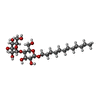 ChemComp-LMT: |
-Macromolecule #3: 1,2-DIACYL-SN-GLYCERO-3-PHOSPHOCHOLINE
| Macromolecule | Name: 1,2-DIACYL-SN-GLYCERO-3-PHOSPHOCHOLINE / type: ligand / ID: 3 / Number of copies: 2 / Formula: PC1 |
|---|---|
| Molecular weight | Theoretical: 790.145 Da |
| Chemical component information | 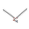 ChemComp-PC1: |
-Macromolecule #4: 1,2-DIACYL-SN-GLYCERO-3-PHOSHOCHOLINE
| Macromolecule | Name: 1,2-DIACYL-SN-GLYCERO-3-PHOSHOCHOLINE / type: ligand / ID: 4 / Number of copies: 2 / Formula: PCF |
|---|---|
| Molecular weight | Theoretical: 734.039 Da |
| Chemical component information | 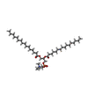 ChemComp-PCF: |
-Macromolecule #5: Dipalmitoyl Phosphatidylinositol 3,5-bisphosphate
| Macromolecule | Name: Dipalmitoyl Phosphatidylinositol 3,5-bisphosphate / type: ligand / ID: 5 / Number of copies: 2 / Formula: A1JDN |
|---|---|
| Molecular weight | Theoretical: 970.992 Da |
-Experimental details
-Structure determination
| Method | cryo EM |
|---|---|
 Processing Processing | single particle reconstruction |
| Aggregation state | particle |
-Sample preparation
| Concentration | 5 mg/mL | ||||||||||||||||||
|---|---|---|---|---|---|---|---|---|---|---|---|---|---|---|---|---|---|---|---|
| Buffer | pH: 7.5 Component:
Details: 20 mM HEPES pH 7.5; 200mM NaCl; 0.015% DDM / 0.0015% CHS; 0.64mM PI(3,5)P2 (16:0) | ||||||||||||||||||
| Grid | Model: Quantifoil R1.2/1.3 / Material: GOLD / Mesh: 300 / Support film - Material: CARBON / Support film - topology: HOLEY / Pretreatment - Type: GLOW DISCHARGE / Pretreatment - Time: 60 sec. / Pretreatment - Atmosphere: AIR | ||||||||||||||||||
| Vitrification | Cryogen name: ETHANE / Chamber humidity: 100 % / Chamber temperature: 277 K / Instrument: FEI VITROBOT MARK IV Details: Sample vol 3ul; blot force -5; Blot time 9s; Wait time 20sec. |
- Electron microscopy
| Microscope | TFS KRIOS |
|---|---|
| Specialist optics | Energy filter - Name: TFS Selectris X / Energy filter - Slit width: 10 eV |
| Image recording | Film or detector model: TFS FALCON 4i (4k x 4k) / Number grids imaged: 1 / Number real images: 9002 / Average exposure time: 4.7 sec. / Average electron dose: 50.0 e/Å2 |
| Electron beam | Acceleration voltage: 300 kV / Electron source:  FIELD EMISSION GUN FIELD EMISSION GUN |
| Electron optics | C2 aperture diameter: 50.0 µm / Illumination mode: FLOOD BEAM / Imaging mode: BRIGHT FIELD / Nominal defocus max: 2.2 µm / Nominal defocus min: 0.8 µm / Nominal magnification: 130000 |
| Sample stage | Specimen holder model: FEI TITAN KRIOS AUTOGRID HOLDER / Cooling holder cryogen: NITROGEN |
| Experimental equipment |  Model: Titan Krios / Image courtesy: FEI Company |
+Image processing

-Atomic model buiding 1
| Initial model | PDB ID: Chain - Source name: AlphaFold / Chain - Initial model type: in silico model |
|---|---|
| Details | Initial model fitted and manually rebuilt/refined in COOT and final refinement in ISOLDE and PHENIX |
| Refinement | Space: REAL / Protocol: FLEXIBLE FIT |
| Output model | PDB-9r8m: |

