 Movie
Movie Controller

[English] 日本語
 Yorodumi
- EMDB-53357: Cryo-electron tomograms of cryo-FIB milled E. coli lacking LpoB. -
+ Open data

- Basic information
| Entry |  | ||||||||||||||||||
|---|---|---|---|---|---|---|---|---|---|---|---|---|---|---|---|---|---|---|---|
| Title | Cryo-electron tomograms of cryo-FIB milled E. coli lacking LpoB. | ||||||||||||||||||
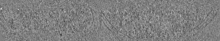
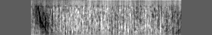
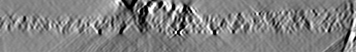
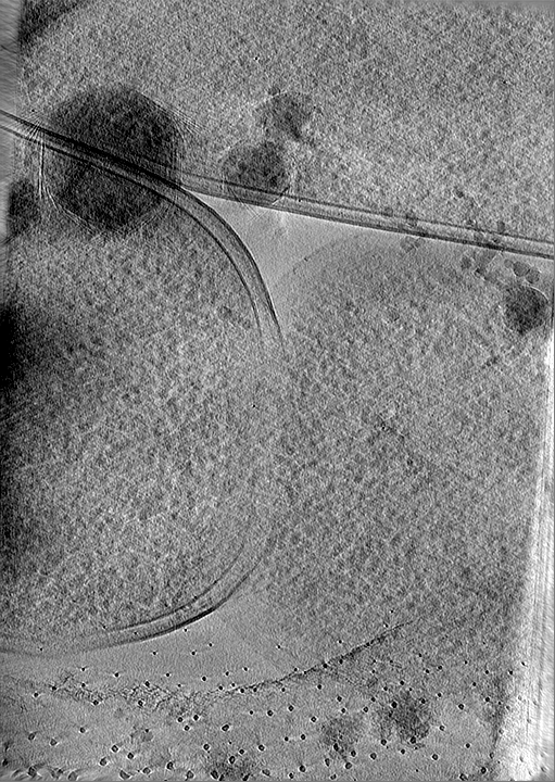
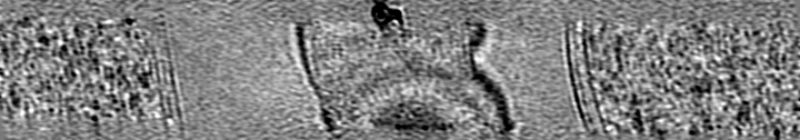
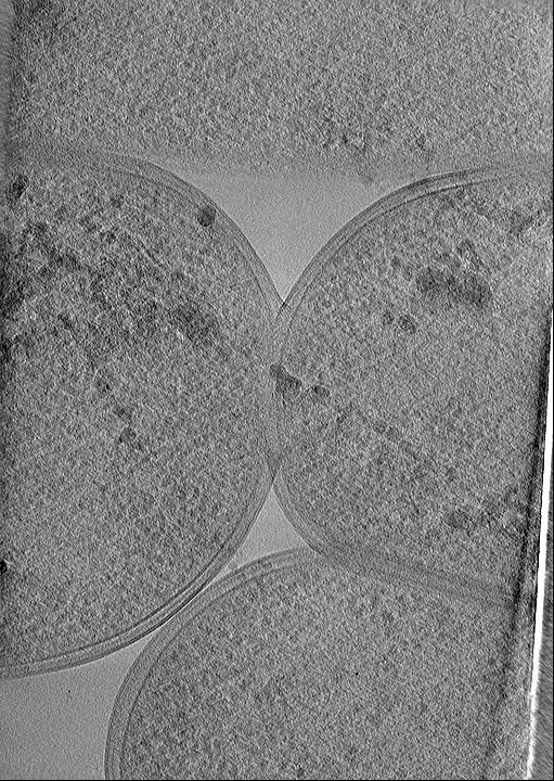
 Map data Map data | Dividing E.coli cell lacking LpoB. Septation stage. | ||||||||||||||||||
 Sample Sample |
| ||||||||||||||||||
 Keywords Keywords | PBP1b / bacteria / division / peptidoglycan / CELL CYCLE | ||||||||||||||||||
| Biological species |  | ||||||||||||||||||
| Method | electron tomography / cryo EM | ||||||||||||||||||
 Authors Authors | Navarro PP / Bernhardt TG | ||||||||||||||||||
| Funding support | European Union,  Switzerland, Switzerland,  United States, 5 items United States, 5 items
| ||||||||||||||||||
 Citation Citation | Journal: bioRxiv / Year: 2025 Title: The aPBP-type cell wall synthase PBP1b plays a specialized role in fortifying the division site against osmotic rupture. Authors: Paula P Navarro / Andrea Vettiger / Roman Hajdu / Virly Y Ananda / Alejandro López-Tavares / Ernst W Schmid / Johannes C Walter / Martin Loose / Luke H Chao / Thomas G Bernhardt Abstract: A multi-protein system called the divisome promotes bacterial division. This apparatus synthesizes the peptidoglycan (PG) cell wall layer that forms the daughter cell poles and protects them from ...A multi-protein system called the divisome promotes bacterial division. This apparatus synthesizes the peptidoglycan (PG) cell wall layer that forms the daughter cell poles and protects them from osmotic lysis. In the model Gram-negative bacterium , PG synthases called class A penicillin-binding proteins (aPBPs) have been proposed to play crucial roles in division. However, there is limited experimental support for aPBPs playing a specialized role in division that is distinct from their general function in the expansion and fortification of the PG matrix. Here, we present cryogenic electron tomography data indicating that the aPBP-type enzyme PBP1b is required to produce a wedge-like density of PG at the division site. Furthermore, atomic force and live cell microscopy showed that loss of this structure weakens the division site and renders it susceptible to lysis. Surprisingly, we found that the lipoprotein activator LpoB needed to promote the general function of PBP1b was not required for normal division site architecture or its integrity. Additionally, we show that of the two PBP1b isoforms produced in cells, it is the one with an extended cytoplasmic N-terminus that functions in division, likely via recruitment by the FtsA component of the divisome. Altogether, our results demonstrate that PBP1b plays a specialized, LpoB-independent role in cell division involving the biogenesis of a PG structure that prevents osmotic rupture. The conservation of aPBPs with extended cytoplasmic N-termini suggests that other Gram-negative bacteria may use similar mechanisms to reinforce their division site. | ||||||||||||||||||
| History |
|
- Structure visualization
| Supplemental images |
|---|

- Downloads & links
-EMDB archive
| Map data | emd_53357.map.gz | 16.3 MB |  EMDB map data format EMDB map data format | |
|---|---|---|---|---|
| Header (meta data) | emd-53357-v30.xmlemd-53357.xml | 32.4 KB 32.4 KB | Display Display | EMDB header |
| Images |  emd_53357.png emd_53357.png | 193.1 KB | ||
| Filedesc metadata | emd-53357.cif.gz | 5.4 KB | ||
| Others | emd_53357_additional_1.map.gzemd_53357_additional_10.map.gzemd_53357_additional_2.map.gzemd_53357_additional_3.map.gzemd_53357_additional_4.map.gzemd_53357_additional_5.map.gzemd_53357_additional_6.map.gzemd_53357_additional_7.map.gzemd_53357_additional_8.map.gzemd_53357_additional_9.map.gz | 7 MB 22.1 MB 155.9 MB 21.9 MB 96.5 MB 159 MB 52.7 MB 10.4 MB 138.5 MB 64.4 MB | ||
| Archive directory |  http://ftp.pdbj.org/pub/emdb/structures/EMD-53357ftp://ftp.pdbj.org/pub/emdb/structures/EMD-53357 http://ftp.pdbj.org/pub/emdb/structures/EMD-53357ftp://ftp.pdbj.org/pub/emdb/structures/EMD-53357 | HTTPS FTP |
-Related structure data


-Links
| EMDB pages | EMDB (EBI/PDBe) / EMDataResource |
|---|
-Map
| File | Download / File: emd_53357.map.gz / Format: CCP4 / Size: 30.5 MB / Type: IMAGE STORED AS SIGNED BYTE | ||||||||||||||||||||||||||||||||
|---|---|---|---|---|---|---|---|---|---|---|---|---|---|---|---|---|---|---|---|---|---|---|---|---|---|---|---|---|---|---|---|---|---|
| Annotation | Dividing E.coli cell lacking LpoB. Septation stage. | ||||||||||||||||||||||||||||||||
| Projections & slices | Image control
Images are generated by Spider. generated in cubic-lattice coordinate | ||||||||||||||||||||||||||||||||
| Voxel size | X=Y=Z: 21.64 Å | ||||||||||||||||||||||||||||||||
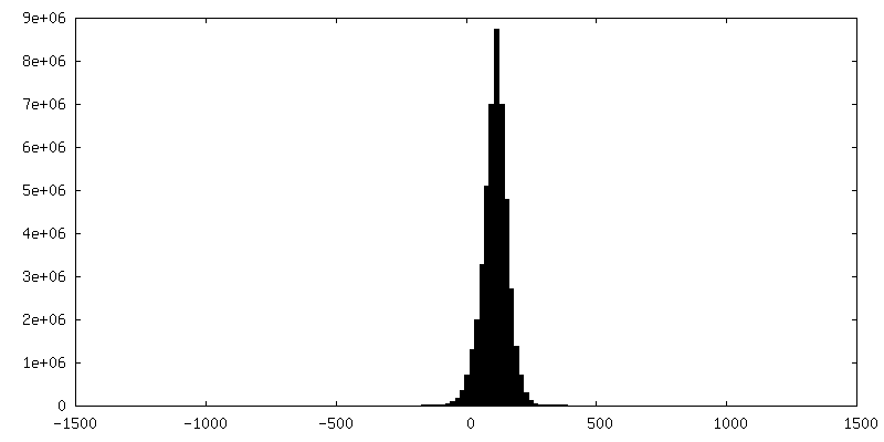
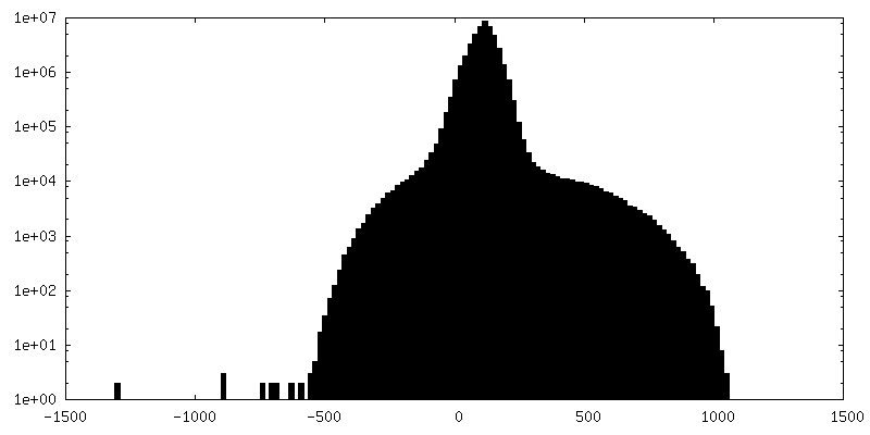
| Density |
| ||||||||||||||||||||||||||||||||
| Symmetry | Space group: 1 | ||||||||||||||||||||||||||||||||
| Details | EMDB XML:
|
 Z (Sec.)
 Y (Row.)
 X (Col.)


-Supplemental data
+Additional map: Dividing E.coli cell lacking LpoB. Septation stage. Low-passed.


+Additional map: Dividing E.coli cell lacking LpoB. Cytokinesis stage.


+Additional map: Dividing E.coli cell lacking LpoB. Constriction stage. Low-passed.


+Additional map: Dividing E.coli cell lacking LpoB. Constriction stage.


+Additional map: Dividing E.coli cell lacking LpoB. Constriction stage. Low-passed.


+Additional map: Dividing E.coli cell lacking LpoB. Septation stage.


+Additional map: Dividing E.coli cell lacking LpoB. Septation stage. Low-passed.


+Additional map: Dividing E.coli cell lacking LpoB. Constriction stage.


+Additional map: Pole of E.coli lacking LpoB.


+Additional map: Dividing E.coli cell lacking LpoB. Septation stage.


- Sample components
-Entire : Dividing E. coli cells lacking LpoB
| Entire | Name: Dividing E. coli cells lacking LpoB |
|---|---|
| Components |
|
-Supramolecule #1: Dividing E. coli cells lacking LpoB
| Supramolecule | Name: Dividing E. coli cells lacking LpoB / type: cell / ID: 1 / Parent: 0 |
|---|---|
| Source (natural) | Organism: |
-Experimental details
-Structure determination
| Method | cryo EM |
|---|---|
 Processing Processing | electron tomography |
| Aggregation state | cell |
-Sample preparation
| Buffer | pH: 7.5 / Details: LB media |
|---|---|
| Grid | Model: C-flat-2/1 / Material: COPPER / Mesh: 200 / Support film - Material: CARBON / Support film - topology: HOLEY / Support film - Film thickness: 20 / Pretreatment - Type: GLOW DISCHARGE / Pretreatment - Time: 30 sec. / Pretreatment - Atmosphere: AIR / Details: glow discharged for 30 seconds at 15 mA |
| Vitrification | Cryogen name: ETHANE / Chamber humidity: 100 % / Chamber temperature: 293 K / Instrument: FEI VITROBOT MARK IV Details: One-side blotting time of 13 seconds and blotting force of 10. Customized parafilm sheets were used for one-side blotting.. |
| Details | Cells grown to O.D600 = 0.3. |
| Sectioning | Focused ion beam - Instrument: OTHER / Focused ion beam - Ion: OTHER / Focused ion beam - Voltage: 30 / Focused ion beam - Current: 0.1 / Focused ion beam - Duration: 1200 / Focused ion beam - Temperature: 83 K / Focused ion beam - Initial thickness: 1200 / Focused ion beam - Final thickness: 200 Focused ion beam - Details: The value given for _em_focused_ion_beam.instrument is Aquilos 2. This is not in a list of allowed values {'OTHER', 'DB235'} so OTHER is written into the XML file. |
- Electron microscopy
| Microscope | TFS KRIOS |
|---|---|
| Specialist optics | Energy filter - Name: GIF Bioquantum |
| Image recording | Film or detector model: GATAN K3 BIOQUANTUM (6k x 4k) / Number grids imaged: 6 / Average electron dose: 3.0 e/Å2 |
| Electron beam | Acceleration voltage: 300 kV / Electron source:  FIELD EMISSION GUN FIELD EMISSION GUN |
| Electron optics | Calibrated defocus max: 7.0 µm / Calibrated defocus min: 1.0 µm / Calibrated magnification: 33000 / Illumination mode: FLOOD BEAM / Imaging mode: BRIGHT FIELD / Cs: 2.7 mm / Nominal defocus max: 5.0 µm / Nominal defocus min: 3.0 µm / Nominal magnification: 33000 |
| Sample stage | Specimen holder model: FEI TITAN KRIOS AUTOGRID HOLDER / Cooling holder cryogen: NITROGEN |
| Experimental equipment |  Model: Titan Krios / Image courtesy: FEI Company |
-Image processing
| Final reconstruction | Software - Name: IMOD / Number images used: 40 |
|---|
