 ムービー
ムービー コントローラー


+ データを開く

- 基本情報
基本情報
| 登録情報 | データベース: EMDB / ID: EMD-5330 | |||||||||
|---|---|---|---|---|---|---|---|---|---|---|
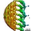
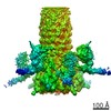
| タイトル | Cryo-electron tomography reveals novel interactions and doublet-specific structures in the I1 dynein | |||||||||
 マップデータ マップデータ | This is a subtomogram average of the I1 inner dynein complex in wild type Chlamydomonas flagella | |||||||||
 試料 試料 |
| |||||||||
 キーワード キーワード | axoneme / molecular motor / motility regulation / flagella | |||||||||
| 生物種 |   Chlamydomonas reinhardtii (クラミドモナス) Chlamydomonas reinhardtii (クラミドモナス) | |||||||||
| 手法 | サブトモグラム平均法 / クライオ電子顕微鏡法 / 解像度: 39.0 Å | |||||||||
 データ登録者 データ登録者 | Heuser T / Barber CF / Lin J / Krell J / Rebesco M / Porter ME / Nicastro D | |||||||||
 引用 引用 | ジャーナル: Proc Natl Acad Sci U S A / 年: 2012 タイトル: Cryoelectron tomography reveals doublet-specific structures and unique interactions in the I1 dynein. 著者: Thomas Heuser / Cynthia F Barber / Jianfeng Lin / Jeremy Krell / Matthew Rebesco / Mary E Porter / Daniela Nicastro /  要旨: Cilia and flagella are highly conserved motile and sensory organelles in eukaryotes, and defects in ciliary assembly and motility cause many ciliopathies. The two-headed I1 inner arm dynein is a ...Cilia and flagella are highly conserved motile and sensory organelles in eukaryotes, and defects in ciliary assembly and motility cause many ciliopathies. The two-headed I1 inner arm dynein is a critical regulator of ciliary and flagellar beating. To understand I1 architecture and function better, we analyzed the 3D structure and composition of the I1 dynein in Chlamydomonas axonemes by cryoelectron tomography and subtomogram averaging. Our data revealed several connections from the I1 dynein to neighboring structures that are likely to be important for assembly and/or regulation, including a tether linking one I1 motor domain to the doublet microtubule and doublet-specific differences potentially contributing to the asymmetrical distribution of dynein activity required for ciliary beating. We also imaged three I1 mutants and analyzed their polypeptide composition using 2D gel-based proteomics. Structural and biochemical comparisons revealed the likely location of the regulatory IC138 phosphoprotein and its associated subcomplex. Overall, our studies demonstrate that I1 dynein is connected to multiple structures within the axoneme, and therefore ideally positioned to integrate signals that regulate ciliary motility. | |||||||||
| 履歴 |
|
- 構造の表示
構造の表示
| ムービー |
 ムービービューア ムービービューア |
|---|---|
| 構造ビューア | EMマップ: SurfViewMolmilJmol/JSmol |
| 添付画像 |

- ダウンロードとリンク
-EMDBアーカイブ
| マップデータ | emd_5330.map.gz | 284.5 KB | EMDBマップデータ形式 | |
|---|---|---|---|---|
| ヘッダ (付随情報) | emd-5330-v30.xmlemd-5330.xml | 10.1 KB 10.1 KB | 表示 表示 | EMDBヘッダ |
| 画像 | emd_5330_1.tif | 184.9 KB | ||
| アーカイブディレクトリ |  http://ftp.pdbj.org/pub/emdb/structures/EMD-5330ftp://ftp.pdbj.org/pub/emdb/structures/EMD-5330 http://ftp.pdbj.org/pub/emdb/structures/EMD-5330ftp://ftp.pdbj.org/pub/emdb/structures/EMD-5330 | HTTPS FTP |
-検証レポート
| 文書・要旨 | emd_5330_validation.pdf.gz | 78.3 KB | 表示 | EMDB検証レポート |
|---|---|---|---|---|
| 文書・詳細版 | emd_5330_full_validation.pdf.gz | 77.4 KB | 表示 | |
| XML形式データ | emd_5330_validation.xml.gz | 493 B | 表示 | |
| アーカイブディレクトリ | https://ftp.pdbj.org/pub/emdb/validation_reports/EMD-5330ftp://ftp.pdbj.org/pub/emdb/validation_reports/EMD-5330 | HTTPS FTP |
-関連構造データ
| 類似構造データ |
|---|
-リンク
| EMDBのページ | EMDB (EBI/PDBe) / EMDataResource |
|---|
-マップ
| ファイル | ダウンロード / ファイル: emd_5330.map.gz / 形式: CCP4 / 大きさ: 336.9 KB / タイプ: IMAGE STORED AS FLOATING POINT NUMBER (4 BYTES) | ||||||||||||||||||||||||||||||||||||||||||||||||||||||||||||||||||||
|---|---|---|---|---|---|---|---|---|---|---|---|---|---|---|---|---|---|---|---|---|---|---|---|---|---|---|---|---|---|---|---|---|---|---|---|---|---|---|---|---|---|---|---|---|---|---|---|---|---|---|---|---|---|---|---|---|---|---|---|---|---|---|---|---|---|---|---|---|---|
| 注釈 | This is a subtomogram average of the I1 inner dynein complex in wild type Chlamydomonas flagella | ||||||||||||||||||||||||||||||||||||||||||||||||||||||||||||||||||||
| 投影像・断面図 | 画像のコントロール
画像は Spider により作成 これらの図は立方格子座標系で作成されたものです | ||||||||||||||||||||||||||||||||||||||||||||||||||||||||||||||||||||
| ボクセルのサイズ | X=Y=Z: 9.5 Å | ||||||||||||||||||||||||||||||||||||||||||||||||||||||||||||||||||||
| 密度 |
| ||||||||||||||||||||||||||||||||||||||||||||||||||||||||||||||||||||
| 対称性 | 空間群: 1 | ||||||||||||||||||||||||||||||||||||||||||||||||||||||||||||||||||||
| 詳細 | EMDB XML:
CCP4マップ ヘッダ情報:
| ||||||||||||||||||||||||||||||||||||||||||||||||||||||||||||||||||||
 Z (Sec.)
 Y (Row.)
 X (Col.)


-添付データ
- 試料の構成要素
-全体 : Cryo-electron tomography and subtomographic average (750 axonemal...
| 全体 | 名称: Cryo-electron tomography and subtomographic average (750 axonemal repeats) of isolated axonemes of wild type Chlamydomonas (CC 125, 137c), I1 dynein complex (dynein f) is bound to the doublet ...名称: Cryo-electron tomography and subtomographic average (750 axonemal repeats) of isolated axonemes of wild type Chlamydomonas (CC 125, 137c), I1 dynein complex (dynein f) is bound to the doublet microtubule and is connected to neighboring structures. |
|---|---|
| 要素 |
|
-超分子 #1000: Cryo-electron tomography and subtomographic average (750 axonemal...
| 超分子 | 名称: Cryo-electron tomography and subtomographic average (750 axonemal repeats) of isolated axonemes of wild type Chlamydomonas (CC 125, 137c), I1 dynein complex (dynein f) is bound to the doublet ...名称: Cryo-electron tomography and subtomographic average (750 axonemal repeats) of isolated axonemes of wild type Chlamydomonas (CC 125, 137c), I1 dynein complex (dynein f) is bound to the doublet microtubule and is connected to neighboring structures. タイプ: sample / ID: 1000 / Number unique components: 1 |
|---|
-超分子 #1: I1 dynein complex
| 超分子 | 名称: I1 dynein complex / タイプ: organelle_or_cellular_component / ID: 1 / Name.synonym: dynein f / コピー数: 2 / 組換発現: No / データベース: NCBI |
|---|---|
| 由来(天然) | 生物種: Chlamydomonas reinhardtii (クラミドモナス) 株: CC-125, 137c / 別称: unicellular green algae / 細胞: Chlamydomonas reinhardtii / Organelle: eukaryotic flagella |
-実験情報
-構造解析
| 手法 | クライオ電子顕微鏡法 |
|---|---|
 解析 解析 | サブトモグラム平均法 |
-試料調製
| 濃度 | 1 mg/mL |
|---|---|
| 緩衝液 | pH: 7.4 詳細: 10 mM HEPES, pH 7.4, 25 mM NaCl, 4 mM MgSO4, 1 mM EGTA, 0.1 mM EDTA |
| グリッド | 詳細: Quantifoil holey carbon grids Cu 200 mesh R2/2 |
| 凍結 | 凍結剤: ETHANE / チャンバー内温度: 100 K / 装置: HOMEMADE PLUNGER / 手法: front-side blotting for 2-3 seconds |
- 電子顕微鏡法
| 顕微鏡 | FEI TECNAI F30 |
|---|---|
| 温度 | 平均: 80 K |
| 特殊光学系 | エネルギーフィルター - 名称: GATAN postcolumn filter GIF エネルギーフィルター - エネルギー下限: 0.0 eV エネルギーフィルター - エネルギー上限: 20.0 eV |
| 日付 | 2004年2月20日 |
| 撮影 | カテゴリ: CCD フィルム・検出器のモデル: GENERIC GATAN (2k x 2k) 平均電子線量: 100 e/Å2 |
| 電子線 | 加速電圧: 300 kV / 電子線源:  FIELD EMISSION GUN FIELD EMISSION GUN |
| 電子光学系 | 照射モード: OTHER / 撮影モード: BRIGHT FIELD / 最大 デフォーカス(公称値): 8.0 µm / 最小 デフォーカス(公称値): 6.0 µm / 倍率(公称値): 13500 |
| 試料ステージ | 試料ホルダー: Eucentric / 試料ホルダーモデル: GATAN LIQUID NITROGEN / Tilt series - Axis1 - Min angle: -60 ° / Tilt series - Axis1 - Max angle: 60 ° |
| 実験機器 |  モデル: Tecnai F30 / 画像提供: FEI Company |
-画像解析
| 詳細 | 750 axonemal repeats (96 nm long) from 5 tomograms (reconstructed using fiducial alignment and weighted backprojection, IMOD software, Kremer et al. 1996) were aligned and averaged using the PEET software (bio3d.colorado.edu, Nicastro et al. 2006). Average number of tilts used in the 3D reconstructions: 80. Average tomographic tilt angle increment: 1.5. |
|---|---|
| 最終 再構成 | アルゴリズム: OTHER / 解像度のタイプ: BY AUTHOR / 解像度: 39.0 Å / 解像度の算出法: FSC 0.5 CUT-OFF / ソフトウェア - 名称: IMOD 詳細: Final maps were calculated by averaging 750 particles from 5 tomograms |
