+ Open data
- Basic information
| Entry |  | |||||||||
|---|---|---|---|---|---|---|---|---|---|---|
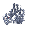
| Title | cryoEM structure of Bovine Serum Albumin | |||||||||
 Map data Map data | Final map from cryoSPARC refinement | |||||||||
 Sample Sample |
| |||||||||
 Keywords Keywords | Lipid binding / metal binding / Albumin / disulfide rich / protein binding / METAL BINDING PROTEIN / TRANSPORT PROTEIN | |||||||||
| Function / homology |  Function and homology information Function and homology informationcellular response to calcium ion starvation / enterobactin binding / negative regulation of mitochondrial depolarization / toxic substance binding / fatty acid binding / cellular response to starvation / pyridoxal phosphate binding / protein-containing complex / extracellular space / DNA binding ...cellular response to calcium ion starvation / enterobactin binding / negative regulation of mitochondrial depolarization / toxic substance binding / fatty acid binding / cellular response to starvation / pyridoxal phosphate binding / protein-containing complex / extracellular space / DNA binding / extracellular region / metal ion binding / cytoplasm Similarity search - Function | |||||||||
| Biological species |  | |||||||||
| Method | single particle reconstruction / cryo EM / Resolution: 3.1 Å | |||||||||
 Authors Authors | Manikandan K / Jason VR | |||||||||
| Funding support |  United Kingdom, 1 items United Kingdom, 1 items
| |||||||||
 Citation Citation |  Journal: J Struct Biol / Year: 2024 Journal: J Struct Biol / Year: 2024Title: The CryoEM structure of human serum albumin in complex with ligands. Authors: Claudio Catalano / Kyle W Lucier / Dennis To / Skerdi Senko / Nhi L Tran / Ashlyn C Farwell / Sabrina M Silva / Phat V Dip / Nicole Poweleit / Giovanna Scapin /  Abstract: Human serum albumin (HSA) is the most prevalent plasma protein in the human body, accounting for 60 % of the total plasma protein. HSA plays a major pharmacokinetic function, serving as a ...Human serum albumin (HSA) is the most prevalent plasma protein in the human body, accounting for 60 % of the total plasma protein. HSA plays a major pharmacokinetic function, serving as a facilitator in the distribution of endobiotics and xenobiotics within the organism. In this paper we report the cryoEM structures of HSA in the apo form and in complex with two ligands (salicylic acid and teniposide) at a resolution of 3.5, 3.7 and 3.4 Å, respectively. We expand upon previously published work and further demonstrate that sub-4 Å maps of ∼60 kDa proteins can be routinely obtained using a 200 kV microscope, employing standard workflows. Most importantly, these maps allowed for the identification of small molecule ligands, emphasizing the practical applicability of this methodology and providing a starting point for subsequent computational modeling and in silico optimization. | |||||||||
| History |
|
- Structure visualization
| Supplemental images |
|---|
- Downloads & links
-EMDB archive
| Map data |  emd_53298.map.gz emd_53298.map.gz | 32 MB |  EMDB map data format EMDB map data format | |
|---|---|---|---|---|
| Header (meta data) |  emd-53298-v30.xml emd-53298-v30.xml emd-53298.xml emd-53298.xml | 22.8 KB 22.8 KB | Display Display |  EMDB header EMDB header |
| Images |  emd_53298.png emd_53298.png | 35.9 KB | ||
| Masks |  emd_53298_msk_1.map emd_53298_msk_1.map | 64 MB |  Mask map Mask map | |
| Filedesc metadata |  emd-53298.cif.gz emd-53298.cif.gz | 6.5 KB | ||
| Others |  emd_53298_additional_1.map.gz emd_53298_additional_1.map.gz emd_53298_additional_2.map.gz emd_53298_additional_2.map.gz emd_53298_half_map_1.map.gz emd_53298_half_map_1.map.gz emd_53298_half_map_2.map.gz emd_53298_half_map_2.map.gz | 31.8 MB 32.1 MB 59.3 MB 59.3 MB | ||
| Archive directory |  http://ftp.pdbj.org/pub/emdb/structures/EMD-53298 http://ftp.pdbj.org/pub/emdb/structures/EMD-53298 ftp://ftp.pdbj.org/pub/emdb/structures/EMD-53298 ftp://ftp.pdbj.org/pub/emdb/structures/EMD-53298 | HTTPS FTP |
-Validation report
| Summary document |  emd_53298_validation.pdf.gz emd_53298_validation.pdf.gz | 724.9 KB | Display |  EMDB validaton report EMDB validaton report |
|---|---|---|---|---|
| Full document |  emd_53298_full_validation.pdf.gz emd_53298_full_validation.pdf.gz | 724.5 KB | Display | |
| Data in XML |  emd_53298_validation.xml.gz emd_53298_validation.xml.gz | 12.2 KB | Display | |
| Data in CIF |  emd_53298_validation.cif.gz emd_53298_validation.cif.gz | 14.5 KB | Display | |
| Arichive directory |  https://ftp.pdbj.org/pub/emdb/validation_reports/EMD-53298 https://ftp.pdbj.org/pub/emdb/validation_reports/EMD-53298 ftp://ftp.pdbj.org/pub/emdb/validation_reports/EMD-53298 ftp://ftp.pdbj.org/pub/emdb/validation_reports/EMD-53298 | HTTPS FTP |
-Related structure data
| Related structure data |  9qqdMC M: atomic model generated by this map C: citing same article ( |
|---|---|
| Similar structure data | Similarity search - Function & homology  F&H Search F&H Search |
- Links
Links
| EMDB pages |  EMDB (EBI/PDBe) / EMDB (EBI/PDBe) /  EMDataResource EMDataResource |
|---|---|
| Related items in Molecule of the Month |
- Map
Map
| File |  Download / File: emd_53298.map.gz / Format: CCP4 / Size: 64 MB / Type: IMAGE STORED AS FLOATING POINT NUMBER (4 BYTES) Download / File: emd_53298.map.gz / Format: CCP4 / Size: 64 MB / Type: IMAGE STORED AS FLOATING POINT NUMBER (4 BYTES) | ||||||||||||||||||||||||||||||||||||
|---|---|---|---|---|---|---|---|---|---|---|---|---|---|---|---|---|---|---|---|---|---|---|---|---|---|---|---|---|---|---|---|---|---|---|---|---|---|
| Annotation | Final map from cryoSPARC refinement | ||||||||||||||||||||||||||||||||||||
| Projections & slices | Image control
Images are generated by Spider. | ||||||||||||||||||||||||||||||||||||
| Voxel size | X=Y=Z: 0.925 Å | ||||||||||||||||||||||||||||||||||||
| Density |
| ||||||||||||||||||||||||||||||||||||
| Symmetry | Space group: 1 | ||||||||||||||||||||||||||||||||||||
| Details | EMDB XML:
|
-Supplemental data
-Mask #1
| File |  emd_53298_msk_1.map emd_53298_msk_1.map | ||||||||||||
|---|---|---|---|---|---|---|---|---|---|---|---|---|---|
| Projections & Slices |
| ||||||||||||
| Density Histograms |
-Additional map: focused mask refinement map
| File | emd_53298_additional_1.map | ||||||||||||
|---|---|---|---|---|---|---|---|---|---|---|---|---|---|
| Annotation | focused mask refinement map | ||||||||||||
| Projections & Slices |
| ||||||||||||
| Density Histograms |
-Additional map: sharpened combined map
| File | emd_53298_additional_2.map | ||||||||||||
|---|---|---|---|---|---|---|---|---|---|---|---|---|---|
| Annotation | sharpened combined map | ||||||||||||
| Projections & Slices |
| ||||||||||||
| Density Histograms |
-Half map: Half map 1
| File | emd_53298_half_map_1.map | ||||||||||||
|---|---|---|---|---|---|---|---|---|---|---|---|---|---|
| Annotation | Half map 1 | ||||||||||||
| Projections & Slices |
| ||||||||||||
| Density Histograms |
-Half map: Half map 2
| File | emd_53298_half_map_2.map | ||||||||||||
|---|---|---|---|---|---|---|---|---|---|---|---|---|---|
| Annotation | Half map 2 | ||||||||||||
| Projections & Slices |
| ||||||||||||
| Density Histograms |
- Sample components
-Entire : BSA
| Entire | Name: BSA |
|---|---|
| Components |
|
-Supramolecule #1: BSA
| Supramolecule | Name: BSA / type: organelle_or_cellular_component / ID: 1 / Parent: 0 / Macromolecule list: all |
|---|---|
| Source (natural) | Organism:  |
| Molecular weight | Theoretical: 66 kDa/nm |
-Macromolecule #1: Albumin
| Macromolecule | Name: Albumin / type: protein_or_peptide / ID: 1 / Number of copies: 1 / Enantiomer: LEVO |
|---|---|
| Source (natural) | Organism:  |
| Molecular weight | Theoretical: 65.729984 KDa |
| Recombinant expression | Organism:  |
| Sequence | String: GKSEIAHRFK DLGEEHFKGL VLIAFSQYLQ QCPFDEHVKL VNELTEFAKT CVADESHAGC EKSLHTLFGD ELCKVASLRE TVGDMADCC EKQEPERNEC FLSHKDDSPD LPKLKPDPNT LCDEFKADEK KFWGKYLYEI ARRHPYFYAP ELLYYANKYN G VFQECCQA ...String: GKSEIAHRFK DLGEEHFKGL VLIAFSQYLQ QCPFDEHVKL VNELTEFAKT CVADESHAGC EKSLHTLFGD ELCKVASLRE TVGDMADCC EKQEPERNEC FLSHKDDSPD LPKLKPDPNT LCDEFKADEK KFWGKYLYEI ARRHPYFYAP ELLYYANKYN G VFQECCQA EDKGACVLPK IETMREKVLV SSARQRLRCA SIQKFGERAL KAWSVARLSQ KFPKAEFVEV TKLVTELTKV HK ECCHGDL LECADDRADL AKYICDNQDT ISSKLKECCD KPLLEKSHCI AEVEKDAVPE NLPPLTADFA EDKDVCKNYQ EAK DAFIGS FLYEYSRRHP EYAVSVLLRL AKEYEATLEE CCAKDDPHAC YSTVFDKLKH LVDEPQNLVK QNCDQFEKLG EYGF QNALV VRYTRKVPQV SSPTLVEVSR SLGKVGTRCC TKPESERMPC TEDYLSLILN RLCVLHEKTP VSEKVTKCCT ESLVN RRPC FSALTPDETY VPKAADEKLF TFHADICTAA DTAKQIKKQT ASVELLKHKP KATEEQLKTV MENFVAFVDK CCAADD KEA CFAVEGPKLV VSTQTA UniProtKB: Albumin |
-Experimental details
-Structure determination
| Method | cryo EM |
|---|---|
 Processing Processing | single particle reconstruction |
| Aggregation state | particle |
- Sample preparation
| Concentration | 4 mg/mL |
|---|---|
| Buffer | pH: 7.4 |
| Grid | Model: UltrAuFoil R1.2/1.3 / Material: GOLD / Mesh: 300 |
| Vitrification | Cryogen name: ETHANE |
| Details | sigma A7906 |
- Electron microscopy
| Microscope | TFS KRIOS |
|---|---|
| Image recording | Film or detector model: FEI FALCON IV (4k x 4k) / Average electron dose: 40.0 e/Å2 |
| Electron beam | Acceleration voltage: 300 kV / Electron source:  FIELD EMISSION GUN FIELD EMISSION GUN |
| Electron optics | Illumination mode: FLOOD BEAM / Imaging mode: BRIGHT FIELD / Nominal defocus max: 2.0 µm / Nominal defocus min: 1.0 µm / Nominal magnification: 130000 |
| Sample stage | Specimen holder model: FEI TITAN KRIOS AUTOGRID HOLDER / Cooling holder cryogen: NITROGEN |
| Experimental equipment |  Model: Titan Krios / Image courtesy: FEI Company |
 Movie
Movie Controller


 Z (Sec.)
 Y (Row.)
 X (Col.)


