 Movie
Movie Controller


+ Open data

- Basic information
| Entry |  | |||||||||||||||
|---|---|---|---|---|---|---|---|---|---|---|---|---|---|---|---|---|
| Title | MDA phage capsid | |||||||||||||||
 Map data Map data | ||||||||||||||||
 Sample Sample |
| |||||||||||||||
 Keywords Keywords | bacteriophage / capsid / neisseria / MCP / major capsid protein / filamentous bacteriophage / inovirus / VIRUS | |||||||||||||||
| Function / homology | Integral membrane protein Function and homology information Function and homology information | |||||||||||||||
| Biological species |  Neisseria meningitidis (bacteria) / MDA (bacteria) Neisseria meningitidis (bacteria) / MDA (bacteria) | |||||||||||||||
| Method | helical reconstruction / cryo EM / Resolution: 3.7 Å | |||||||||||||||
 Authors Authors | Boehning J / Bharat TAM | |||||||||||||||
| Funding support |  United Kingdom, 4 items United Kingdom, 4 items
| |||||||||||||||
 Citation Citation | Journal: Proc Natl Acad Sci U S A / Year: 2025 Title: Structure of the virulence-associated filamentous bacteriophage MDAΦ. Authors: Jan Böhning / Miles Graham / Mathieu Coureuil / Abul K Tarafder / Julie Meyer / Xavier Nassif / Emmanuelle Bille / Tanmay A M Bharat /  Abstract: is a human commensal bacterium that can opportunistically invade the bloodstream and cross the blood-brain barrier, where it can cause septicemia and meningitis. These diseases, if left untreated, ... is a human commensal bacterium that can opportunistically invade the bloodstream and cross the blood-brain barrier, where it can cause septicemia and meningitis. These diseases, if left untreated, can be lethal within hours. Hyperinvasive strains often express a genomically encoded filamentous bacteriophage called MDAΦ, which promotes colonization of mucosal host surfaces to facilitate bacterial invasion. How this phage is organized and how it promotes biofilm formation and infection at the molecular level is unclear. Here, we present an electron cryomicroscopy structure of the MDA phage, showing that MDAΦ is a class I filamentous inovirus, with the major capsid protein (MCP) arranged within the phage as a highly curved and densely packed α-helix. Comparison with other filamentous bacteriophages offers clues about inoviral genome encapsidation mechanisms, providing a framework for understanding the evolutionary diversity of inoviruses. A disordered, N-terminal segment in the MCP presents hydrophobic patches on the surface of assembled phage particles, which, together with electron cryotomography data of phage bundles, furnishes a structural rationale for phage-phage interactions that were seen previously in an epithelium adhesion infection model of . Taken together, our results shed light on the structure, organization, and higher-order assembly of a biomedically relevant phage encoded in the genome of a human pathogen. Molecular insights gleaned from this study increase our understanding of phage evolution, phage-mediated bacterial adhesion, and pathogenicity. | |||||||||||||||
| History |
|
- Structure visualization
| Supplemental images |
|---|

- Downloads & links
-EMDB archive
| Map data | emd_53129.map.gz | 226.9 MB | EMDB map data format | |
|---|---|---|---|---|
| Header (meta data) | emd-53129-v30.xmlemd-53129.xml | 19.6 KB 19.6 KB | Display Display | EMDB header |
| FSC (resolution estimation) | emd_53129_fsc.xml | 14.1 KB | Display | FSC data file |
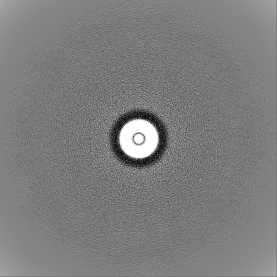
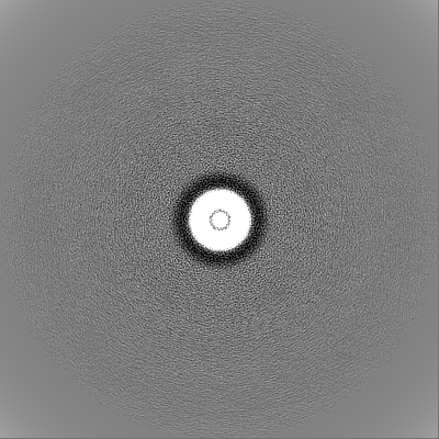
| Images |  emd_53129.png emd_53129.png | 78.5 KB | ||
| Masks | emd_53129_msk_1.map | 244.1 MB | Mask map | |
| Filedesc metadata | emd-53129.cif.gz | 5.7 KB | ||
| Others | emd_53129_half_map_1.map.gzemd_53129_half_map_2.map.gz | 195.5 MB 195.5 MB | ||
| Archive directory |  http://ftp.pdbj.org/pub/emdb/structures/EMD-53129ftp://ftp.pdbj.org/pub/emdb/structures/EMD-53129 http://ftp.pdbj.org/pub/emdb/structures/EMD-53129ftp://ftp.pdbj.org/pub/emdb/structures/EMD-53129 | HTTPS FTP |
-Related structure data
| Related structure data |  9qg9MC M: atomic model generated by this map C: citing same article ( |
|---|---|
| Similar structure data |
-Links
| EMDB pages | EMDB (EBI/PDBe) / EMDataResource |
|---|
-Map
| File | Download / File: emd_53129.map.gz / Format: CCP4 / Size: 244.1 MB / Type: IMAGE STORED AS FLOATING POINT NUMBER (4 BYTES) | ||||||||||||||||||||||||||||||||||||
|---|---|---|---|---|---|---|---|---|---|---|---|---|---|---|---|---|---|---|---|---|---|---|---|---|---|---|---|---|---|---|---|---|---|---|---|---|---|
| Projections & slices | Image control
Images are generated by Spider. | ||||||||||||||||||||||||||||||||||||
| Voxel size | X=Y=Z: 1.236 Å | ||||||||||||||||||||||||||||||||||||
| Density |
| ||||||||||||||||||||||||||||||||||||
| Symmetry | Space group: 1 | ||||||||||||||||||||||||||||||||||||
| Details | EMDB XML:
|
 Z (Sec.)
 Y (Row.)
 X (Col.)


-Supplemental data
-Mask #1
| File | emd_53129_msk_1.map | ||||||||||||
|---|---|---|---|---|---|---|---|---|---|---|---|---|---|
| Projections & Slices |
| ||||||||||||
| Density Histograms |


-Half map: #2
| File | emd_53129_half_map_1.map | ||||||||||||
|---|---|---|---|---|---|---|---|---|---|---|---|---|---|
| Projections & Slices |
| ||||||||||||
| Density Histograms |


-Half map: #1
| File | emd_53129_half_map_2.map | ||||||||||||
|---|---|---|---|---|---|---|---|---|---|---|---|---|---|
| Projections & Slices |
| ||||||||||||
| Density Histograms |


- Sample components
-Entire : MDA
| Entire | Name: MDA (bacteria) |
|---|---|
| Components |
|
-Supramolecule #1: MDA
| Supramolecule | Name: MDA / type: virus / ID: 1 / Parent: 0 / Macromolecule list: all / NCBI-ID: 487 / Sci species name: MDA / Virus type: VIRION / Virus isolate: OTHER / Virus enveloped: No / Virus empty: No |
|---|---|
| Host (natural) | Organism: Neisseria meningitidis (bacteria) / Strain: Z5463 |
-Macromolecule #1: Integral membrane protein
| Macromolecule | Name: Integral membrane protein / type: protein_or_peptide / ID: 1 / Number of copies: 1 / Enantiomer: LEVO |
|---|---|
| Source (natural) | Organism: Neisseria meningitidis (bacteria) / Strain: Z5463 |
| Molecular weight | Theoretical: 5.235297 KDa |
| Sequence | String: DGFDAAAIGT QVANVIMGFV AMVSAVGMAA ITVILAIQGF KMAWSMIKSV K UniProtKB: Integral membrane protein |
-Experimental details
-Structure determination
| Method | cryo EM |
|---|---|
 Processing Processing | helical reconstruction |
| Aggregation state | helical array |
-Sample preparation
| Buffer | pH: 7.4 / Details: PBS |
|---|---|
| Grid | Model: Quantifoil / Material: GOLD / Mesh: 400 / Support film - Material: CARBON / Support film - topology: HOLEY ARRAY / Pretreatment - Type: GLOW DISCHARGE |
| Vitrification | Cryogen name: ETHANE |
- Electron microscopy
| Microscope | TFS KRIOS |
|---|---|
| Image recording | Film or detector model: TFS FALCON 4i (4k x 4k) / Average electron dose: 41.0 e/Å2 |
| Electron beam | Acceleration voltage: 300 kV / Electron source:  FIELD EMISSION GUN FIELD EMISSION GUN |
| Electron optics | Illumination mode: FLOOD BEAM / Imaging mode: BRIGHT FIELD / Nominal defocus max: 2.5 µm / Nominal defocus min: 1.0 µm |
| Sample stage | Specimen holder model: FEI TITAN KRIOS AUTOGRID HOLDER / Cooling holder cryogen: NITROGEN |
| Experimental equipment |  Model: Titan Krios / Image courtesy: FEI Company |
+Image processing

-Atomic model buiding 1
| Details | PHENIX and Servalcat was used |
|---|---|
| Refinement | Space: REAL / Protocol: AB INITIO MODEL |
| Output model | PDB-9qg9: |
