[English] 日本語
Yorodumi
- EMDB-53064: CryoEM structure of transcribing RNA polymerase II elongation com... -

+
Open data


ID or keywords:

Loading...

-
Basic information

Entry
Database: EMDB / ID: EMD-53064
TitleCryoEM structure of transcribing RNA polymerase II elongation complex_3D classification map containing the complete nucleic acid scaffold
Map dataCryoEM structure of transcribing RNA polymerase II elongation complex_3D classification map containing the complete nucleic acid scaffold.
Sample
  • Complex: Yeast RNA polymerase II elongation complex
KeywordsCryoEM / RNA / polymerase / Transcription
Biological speciesSaccharomyces cerevisiae (brewer's yeast)
Methodsingle particle reconstruction / cryo EM / Resolution: 3.12 Å
AuthorsLi Q / Yi G / Zhang P / Wang D
Funding support United Kingdom, United States, 6 items
OrganizationGrant numberCountry
Wellcome Trust206422/Z/17/Z United Kingdom
National Institutes of Health/National Institute of General Medical Sciences (NIH/NIGMS)GM102362 United States
National Institutes of Health/National Institute of General Medical Sciences (NIH/NIGMS)GM148476 United States
National Institutes of Health/National Institute of General Medical Sciences (NIH/NIGMS)GM147652 United States
National Institutes of Health/National Institute of General Medical Sciences (NIH/NIGMS)AI170791-7522 United States
National Institutes of Health/National Institute of General Medical Sciences (NIH/NIGMS)CA280467 United States
Citation
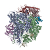
Journal: Mol Cell / Year: 2026
Title: Sub-2 Å cryo-EM structures of transcribing RNA polymerase II reveal critical roles of water molecules in catalysis.
Authors: Qingrong Li / Gangshun Yi / Yue Wu / Sophy Xu / Jenny Chong / Xuhui Huang / Peijun Zhang / Dong Wang /
Abstract: RNA polymerase II (RNA Pol II) is central to gene expression, but its catalytic mechanism remains elusive due to the absence of high-resolution structural data. The role of water molecules in RNA Pol ...RNA polymerase II (RNA Pol II) is central to gene expression, but its catalytic mechanism remains elusive due to the absence of high-resolution structural data. The role of water molecules in RNA Pol II catalysis is unknown. Here, we present 3 high-resolution cryo-electron microscopy structures of active Saccharomyces cerevisiae RNA Pol II elongation complexes in distinct catalytic states: two pre-catalysis states at 1.96 Å and 2.26 Å resolution and a post-catalysis state at 2.33 Å resolution. Each structure contains over 700-1,350 ordered water molecules, many located at functionally critical positions. Comparative analysis shows that these waters play essential roles in proton-transfer steps during RNA Pol II catalysis, facilitating substrate recognition and trigger-loop folding during nucleotide addition. Strikingly, these waters are conserved between prokaryotic and eukaryotic transcription machineries (see Mueller and Darst). These findings provide unprecedented mechanistic insights into RNA Pol II catalysis and reveal vital and evolutionarily conserved roles of water molecules in transcription.
History
DepositionMar 9, 2025-
Header (metadata) releaseMay 13, 2026-
Map releaseMay 13, 2026-
UpdateMay 13, 2026-
Current statusMay 13, 2026Processing site: PDBe / Status: Released

-
Structure visualization

Supplemental images

Downloads & links

-
Map

FileDownload / File: emd_53064.map.gz / Format: CCP4 / Size: 282.6 MB / Type: IMAGE STORED AS FLOATING POINT NUMBER (4 BYTES)
AnnotationCryoEM structure of transcribing RNA polymerase II elongation complex_3D classification map containing the complete nucleic acid scaffold.
Projections & slices

Image control

Size
Brightness
Contrast
Others
AxesZ (Sec.)Y (Row.)X (Col.)
0.83 Å/pix.
x 420 pix.
= 348.18 Å
0.83 Å/pix.
x 420 pix.
= 348.18 Å
0.83 Å/pix.
x 420 pix.
= 348.18 Å

Surface

Projections

Slices (1/3)

Slices (1/2)

Slices (2/3)

Images are generated by Spider.

Voxel sizeX=Y=Z: 0.829 Å
Density
Contour LevelBy AUTHOR: 0.07
Minimum - Maximum-0.19806533 - 0.4467938
Average (Standard dev.)0.000012676724 (±0.016679822)
SymmetrySpace group: 1
Details

EMDB XML:

Map geometry
Axis orderXYZ
Origin000
Dimensions420420420
Spacing420420420
CellA=B=C: 348.18 Å
α=β=γ: 90.0 °

-
Supplemental data

-
Half map: CryoEM structure of transcribing RNA polymerase II elongation...

Fileemd_53064_half_map_1.map
AnnotationCryoEM structure of transcribing RNA polymerase II elongation complex_half A map of 3D classification map containing the complete nucleic acid scaffold.
Projections & Slices
AxesZYX

Projections

Slices (1/2)
Density Histograms

-
Half map: CryoEM structure of transcribing RNA polymerase II elongation...

Fileemd_53064_half_map_2.map
AnnotationCryoEM structure of transcribing RNA polymerase II elongation complex_half B map of 3D classification map containing the complete nucleic acid scaffold.
Projections & Slices
AxesZYX

Projections

Slices (1/2)
Density Histograms

-
Sample components

-
Entire : Yeast RNA polymerase II elongation complex

EntireName: Yeast RNA polymerase II elongation complex
Components
  • Complex: Yeast RNA polymerase II elongation complex

-
Supramolecule #1: Yeast RNA polymerase II elongation complex

SupramoleculeName: Yeast RNA polymerase II elongation complex / type: complex / ID: 1 / Parent: 0
Source (natural)Organism: Saccharomyces cerevisiae (brewer's yeast)
Molecular weightTheoretical: 0.513 kDa/nm

-
Experimental details

-
Structure determination

Methodcryo EM
Processingsingle particle reconstruction
Aggregation stateparticle

-
Sample preparation

Concentration1 mg/mL
BufferpH: 7.5
GridModel: Quantifoil R2/1 / Material: COPPER / Mesh: 300 / Support film - Material: CARBON / Support film - topology: HOLEY
VitrificationCryogen name: ETHANE / Chamber humidity: 100 % / Chamber temperature: 277.15 K / Instrument: FEI VITROBOT MARK II

-
Electron microscopy

MicroscopeTFS KRIOS
Image recordingFilm or detector model: GATAN K3 (6k x 4k) / Average electron dose: 50.0 e/Å2
Electron beamAcceleration voltage: 300 kV / Electron source: FIELD EMISSION GUN
Electron opticsC2 aperture diameter: 100.0 µm / Illumination mode: FLOOD BEAM / Imaging mode: BRIGHT FIELD / Cs: 2.7 mm / Nominal defocus max: 2.5 µm / Nominal defocus min: 0.5 µm
Experimental equipment
Model: Titan Krios / Image courtesy: FEI Company

+
Image processing

CTF correctionType: NONE
Startup modelType of model: PDB ENTRY
PDB model - PDB ID:

Details: 2E2H and 8U9R
Final reconstructionResolution.type: BY AUTHOR / Resolution: 3.12 Å / Resolution method: FSC 0.143 CUT-OFF / Number images used: 50808
Initial angle assignmentType: MAXIMUM LIKELIHOOD
Final angle assignmentType: MAXIMUM LIKELIHOOD
FSC plot (resolution estimation)

+
About Yorodumi

-
News

-
Feb 9, 2022. New format data for meta-information of EMDB entries

New format data for meta-information of EMDB entries

  • Version 3 of the EMDB header file is now the official format.
  • The previous official version 1.9 will be removed from the archive.

Related info.:EMDB header

External links:wwPDB to switch to version 3 of the EMDB data model

-
Aug 12, 2020. Covid-19 info

Covid-19 info

URL: https://pdbj.org/emnavi/covid19.php

New page: Covid-19 featured information page in EM Navigator.

Related info.:Covid-19 info / Mar 5, 2020. Novel coronavirus structure data

+
Mar 5, 2020. Novel coronavirus structure data

Novel coronavirus structure data

Related info.:Yorodumi Speices / Aug 12, 2020. Covid-19 info

External links:COVID-19 featured content - PDBj / Molecule of the Month (242):Coronavirus Proteases

+
Jan 31, 2019. EMDB accession codes are about to change! (news from PDBe EMDB page)

EMDB accession codes are about to change! (news from PDBe EMDB page)

  • The allocation of 4 digits for EMDB accession codes will soon come to an end. Whilst these codes will remain in use, new EMDB accession codes will include an additional digit and will expand incrementally as the available range of codes is exhausted. The current 4-digit format prefixed with “EMD-” (i.e. EMD-XXXX) will advance to a 5-digit format (i.e. EMD-XXXXX), and so on. It is currently estimated that the 4-digit codes will be depleted around Spring 2019, at which point the 5-digit format will come into force.
  • The EM Navigator/Yorodumi systems omit the EMD- prefix.

Related info.:Q: What is EMD? / ID/Accession-code notation in Yorodumi/EM Navigator

External links:EMDB Accession Codes are Changing Soon! / Contact to PDBj

+
Jul 12, 2017. Major update of PDB

Major update of PDB

  • wwPDB released updated PDB data conforming to the new PDBx/mmCIF dictionary.
  • This is a major update changing the version number from 4 to 5, and with Remediation, in which all the entries are updated.
  • In this update, many items about electron microscopy experimental information are reorganized (e.g. em_software).
  • Now, EM Navigator and Yorodumi are based on the updated data.

External links:wwPDB Remediation / Enriched Model Files Conforming to OneDep Data Standards Now Available in the PDB FTP Archive

-
Yorodumi

Thousand views of thousand structures

  • Yorodumi is a browser for structure data from EMDB, PDB, SASBDB, etc.
  • This page is also the successor to EM Navigator detail page, and also detail information page/front-end page for Omokage search.
  • The word "yorodu" (or yorozu) is an old Japanese word meaning "ten thousand". "mi" (miru) is to see.

Related info.:EMDB / PDB / SASBDB / Comparison of 3 databanks / Yorodumi Search / Aug 31, 2016. New EM Navigator & Yorodumi / Yorodumi Papers / Jmol/JSmol / Function and homology information / Changes in new EM Navigator and Yorodumi

Read more