Movie
Movie Controller

[English] 日本語
 Yorodumi
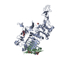
- EMDB-52999: HER2/ErbB2 extracellular domain (ECD) from a near full-length con... -
+ Open data

- Basic information
| Entry |  | |||||||||
|---|---|---|---|---|---|---|---|---|---|---|
| Title | HER2/ErbB2 extracellular domain (ECD) from a near full-length construct solubilized in amphipols. | |||||||||
 Map data Map data | Refined full map (used for model validation | |||||||||
 Sample Sample |
| |||||||||
 Keywords Keywords | Receptor / tyrosine kinase / transmembrane / HER2 / SIGNALING PROTEIN | |||||||||
| Function / homology |  Function and homology information Function and homology informationnegative regulation of immature T cell proliferation in thymus / ERBB3:ERBB2 complex / ERBB2-ERBB4 signaling pathway / immature T cell proliferation in thymus / GRB7 events in ERBB2 signaling / RNA polymerase I core binding / semaphorin receptor complex / Developmental Lineage of Mammary Stem Cells / ErbB-3 class receptor binding / motor neuron axon guidance ...negative regulation of immature T cell proliferation in thymus / ERBB3:ERBB2 complex / ERBB2-ERBB4 signaling pathway / immature T cell proliferation in thymus / GRB7 events in ERBB2 signaling / RNA polymerase I core binding / semaphorin receptor complex / Developmental Lineage of Mammary Stem Cells / ErbB-3 class receptor binding / motor neuron axon guidance / Sema4D induced cell migration and growth-cone collapse / regulation of microtubule-based process / PLCG1 events in ERBB2 signaling / ERBB2-EGFR signaling pathway / enzyme-linked receptor protein signaling pathway / ERBB2 Activates PTK6 Signaling / ERBB2-ERBB3 signaling pathway / Drug-mediated inhibition of ERBB2 signaling / Resistance of ERBB2 KD mutants to trastuzumab / Resistance of ERBB2 KD mutants to sapitinib / Resistance of ERBB2 KD mutants to tesevatinib / Resistance of ERBB2 KD mutants to neratinib / Resistance of ERBB2 KD mutants to osimertinib / Resistance of ERBB2 KD mutants to afatinib / Resistance of ERBB2 KD mutants to AEE788 / Resistance of ERBB2 KD mutants to lapatinib / Drug resistance in ERBB2 TMD/JMD mutants / neurotransmitter receptor localization to postsynaptic specialization membrane / positive regulation of Rho protein signal transduction / positive regulation of MAP kinase activity / oligodendrocyte differentiation / positive regulation of transcription by RNA polymerase I / ERBB2 Regulates Cell Motility / Developmental Lineage of Mammary Gland Myoepithelial Cells / semaphorin-plexin signaling pathway / neuromuscular junction development / PI3K events in ERBB2 signaling / regulation of angiogenesis / positive regulation of protein targeting to membrane / regulation of ERK1 and ERK2 cascade / Schwann cell development / coreceptor activity / Signaling by ERBB2 / TFAP2 (AP-2) family regulates transcription of growth factors and their receptors / myelination / peptidyl-tyrosine phosphorylation / transmembrane receptor protein tyrosine kinase activity / positive regulation of epithelial cell proliferation / GRB2 events in ERBB2 signaling / positive regulation of cell adhesion / SHC1 events in ERBB2 signaling / cell surface receptor protein tyrosine kinase signaling pathway / cellular response to epidermal growth factor stimulus / basal plasma membrane / Constitutive Signaling by Overexpressed ERBB2 / Downregulation of ERBB2:ERBB3 signaling / bioluminescence / positive regulation of translation / generation of precursor metabolites and energy / phosphatidylinositol 3-kinase/protein kinase B signal transduction / neuromuscular junction / wound healing / Signaling by ERBB2 TMD/JMD mutants / receptor protein-tyrosine kinase / Signaling by ERBB2 ECD mutants / Signaling by ERBB2 KD Mutants / cellular response to growth factor stimulus / receptor tyrosine kinase binding / epidermal growth factor receptor signaling pathway / ruffle membrane / Downregulation of ERBB2 signaling / neuron differentiation / Constitutive Signaling by Aberrant PI3K in Cancer / transmembrane signaling receptor activity / PIP3 activates AKT signaling / myelin sheath / heart development / PI5P, PP2A and IER3 Regulate PI3K/AKT Signaling / positive regulation of cell growth / RAF/MAP kinase cascade / protein tyrosine kinase activity / presynaptic membrane / basolateral plasma membrane / protein phosphorylation / positive regulation of MAPK cascade / early endosome / cell population proliferation / cell surface receptor signaling pathway / signaling receptor complex / apical plasma membrane / endosome membrane / intracellular signal transduction / protein heterodimerization activity / signaling receptor binding / negative regulation of apoptotic process / perinuclear region of cytoplasm / signal transduction / nucleoplasm / ATP binding / membrane Similarity search - Function | |||||||||
| Biological species |  Homo sapiens (human) / Homo sapiens (human) /   Aequorea victoria (jellyfish) Aequorea victoria (jellyfish) | |||||||||
| Method | single particle reconstruction / cryo EM / Resolution: 3.77 Å | |||||||||
 Authors Authors | Gragera M / Buschiazzo A / Vacca S | |||||||||
| Funding support | European Union, 1 items
| |||||||||
 Citation Citation | Journal: Sci Adv / Year: 2025 Title: Structural analysis of HER2-trastuzumab complex reveals receptor conformational adaptation. Authors: Santiago Vacca / Marcos Gragera / Alejandro Buschiazzo / David Herreros / James M Krieger / Santiago Bonn-Garcia / Roberto Melero / Carlos Os Sorzano / Jose M Carazo / Ohad Medalia / Andreas Plückthun /    Abstract: Human epidermal growth factor receptor-2 (HER2) is a receptor tyrosine kinase, associated with a variety of malignant tumors, usually through overexpression, resulting in aberrant signaling. ...Human epidermal growth factor receptor-2 (HER2) is a receptor tyrosine kinase, associated with a variety of malignant tumors, usually through overexpression, resulting in aberrant signaling. Trastuzumab (TZB), one of the monoclonal antibodies (mAbs) used in combination with chemotherapy, has become a major therapeutic for HER2-overexpressing cancers. Current structural understanding of HER2 and its interactions with other receptors and with different affinity agents has relied on numerous structures of individual domains of HER2. Here, we subjected purified near full-length HER2 to single-particle cryo-electron microscopy (cryo-EM) analysis. Besides the canonical conformation described in previous structural studies, we report a previously unreported conformation of the HER2 extracellular domain that is stabilized upon TZB binding, which might hamper association with HER3, a receptor with which HER2 forms an oncogenic unit. Together, our findings provide insights into the conformational dynamics of the HER2 receptor and the mechanism of action of TZB. | |||||||||
| History |
|
- Structure visualization
| Supplemental images |
|---|

- Downloads & links
-EMDB archive
| Map data | emd_52999.map.gz | 15.1 MB | EMDB map data format | |
|---|---|---|---|---|
| Header (meta data) | emd-52999-v30.xmlemd-52999.xml | 25.1 KB 25.1 KB | Display Display | EMDB header |
| FSC (resolution estimation) | emd_52999_fsc.xml | 7.1 KB | Display | FSC data file |
| Images |  emd_52999.png emd_52999.png | 81.7 KB | ||
| Masks | emd_52999_msk_1.map | 30.5 MB | Mask map | |
| Filedesc metadata | emd-52999.cif.gz | 7.6 KB | ||
| Others | emd_52999_additional_1.map.gzemd_52999_additional_2.map.gzemd_52999_half_map_1.map.gzemd_52999_half_map_2.map.gz | 27.4 MB 17.1 MB 28.3 MB 28.3 MB | ||
| Archive directory |  http://ftp.pdbj.org/pub/emdb/structures/EMD-52999ftp://ftp.pdbj.org/pub/emdb/structures/EMD-52999 http://ftp.pdbj.org/pub/emdb/structures/EMD-52999ftp://ftp.pdbj.org/pub/emdb/structures/EMD-52999 | HTTPS FTP |
-Related structure data
| Related structure data |  9qbhMC  9qbfC  9qbgC M: atomic model generated by this map C: citing same article ( |
|---|---|
| Similar structure data |


-Links
| EMDB pages | EMDB (EBI/PDBe) / EMDataResource |
|---|---|
| Related items in Molecule of the Month |


-Map
| File | Download / File: emd_52999.map.gz / Format: CCP4 / Size: 30.5 MB / Type: IMAGE STORED AS FLOATING POINT NUMBER (4 BYTES) | ||||||||||||||||||||||||||||||||||||
|---|---|---|---|---|---|---|---|---|---|---|---|---|---|---|---|---|---|---|---|---|---|---|---|---|---|---|---|---|---|---|---|---|---|---|---|---|---|
| Annotation | Refined full map (used for model validation | ||||||||||||||||||||||||||||||||||||
| Projections & slices | Image control
Images are generated by Spider. | ||||||||||||||||||||||||||||||||||||
| Voxel size | X=Y=Z: 1.302 Å | ||||||||||||||||||||||||||||||||||||
| Density |
| ||||||||||||||||||||||||||||||||||||
| Symmetry | Space group: 1 | ||||||||||||||||||||||||||||||||||||
| Details | EMDB XML:
|
 Z (Sec.)
 Y (Row.)
 X (Col.)


-Supplemental data
-Mask #1
| File | emd_52999_msk_1.map | ||||||||||||
|---|---|---|---|---|---|---|---|---|---|---|---|---|---|
| Projections & Slices |
| ||||||||||||
| Density Histograms |


-Additional map: Refined Full map sharpened with deepEMhancer
| File | emd_52999_additional_1.map | ||||||||||||
|---|---|---|---|---|---|---|---|---|---|---|---|---|---|
| Annotation | Refined Full map sharpened with deepEMhancer | ||||||||||||
| Projections & Slices |
| ||||||||||||
| Density Histograms |


-Additional map: Refined Full map sharpened with relion post-process
| File | emd_52999_additional_2.map | ||||||||||||
|---|---|---|---|---|---|---|---|---|---|---|---|---|---|
| Annotation | Refined Full map sharpened with relion post-process | ||||||||||||
| Projections & Slices |
| ||||||||||||
| Density Histograms |


-Half map: #2
| File | emd_52999_half_map_1.map | ||||||||||||
|---|---|---|---|---|---|---|---|---|---|---|---|---|---|
| Projections & Slices |
| ||||||||||||
| Density Histograms |


-Half map: #1
| File | emd_52999_half_map_2.map | ||||||||||||
|---|---|---|---|---|---|---|---|---|---|---|---|---|---|
| Projections & Slices |
| ||||||||||||
| Density Histograms |


- Sample components
-Entire : HER2/ErbB2 extracellular domain (ECD) from a near full-length con...
| Entire | Name: HER2/ErbB2 extracellular domain (ECD) from a near full-length construct solubilized in amphipols. |
|---|---|
| Components |
|
-Supramolecule #1: HER2/ErbB2 extracellular domain (ECD) from a near full-length con...
| Supramolecule | Name: HER2/ErbB2 extracellular domain (ECD) from a near full-length construct solubilized in amphipols. type: complex / ID: 1 / Parent: 0 / Macromolecule list: all Details: The final reconstruction contains only the HER2 ECD. |
|---|---|
| Source (natural) | Organism: Homo sapiens (human) |
| Molecular weight | Theoretical: 112 KDa |
-Macromolecule #1: Receptor tyrosine-protein kinase erbB-2,Green fluorescent protein
| Macromolecule | Name: Receptor tyrosine-protein kinase erbB-2,Green fluorescent protein type: protein_or_peptide / ID: 1 Details: Only the extracellular domain (residues from Q24 to C544) of the molecule was solved in the cryo-EM map,Only the extracellular domain (residues from Q24 to C544) of the molecule was solved ...Details: Only the extracellular domain (residues from Q24 to C544) of the molecule was solved in the cryo-EM map,Only the extracellular domain (residues from Q24 to C544) of the molecule was solved in the cryo-EM map,Only the extracellular domain (residues from Q24 to C544) of the molecule was solved in the cryo-EM map,Only the extracellular domain (residues from Q24 to C544) of the molecule was solved in the cryo-EM map.,Only the extracellular domain (residues from Q24 to C544) of the molecule was solved in the cryo-EM map,Only the extracellular domain (residues from Q24 to C544) of the molecule was solved in the cryo-EM map,Only the extracellular domain (residues from Q24 to C544) of the molecule was solved in the cryo-EM map,Only the extracellular domain (residues from Q24 to C544) of the molecule was solved in the cryo-EM map. Number of copies: 1 / Enantiomer: LEVO / EC number: receptor protein-tyrosine kinase |
|---|---|
| Source (natural) | Organism: Aequorea victoria (jellyfish) |
| Molecular weight | Theoretical: 146.737172 KDa |
| Recombinant expression | Organism:   Spodoptera frugiperda (fall armyworm) Spodoptera frugiperda (fall armyworm) |
| Sequence | String: MKFLVNVALV FMVVYISYIY ADYKDDDDKH HHHHHHHHHL EVLFQGPYPY DVPDYATQVC TGTDMKLRLP ASPETHLDML RHLYQGCQV VQGNLELTYL PTNASLSFLQ DIQEVQGYVL IAHNQVRQVP LQRLRIVRGT QLFEDNYALA VLDNGDPLNN T TPVTGASP ...String: MKFLVNVALV FMVVYISYIY ADYKDDDDKH HHHHHHHHHL EVLFQGPYPY DVPDYATQVC TGTDMKLRLP ASPETHLDML RHLYQGCQV VQGNLELTYL PTNASLSFLQ DIQEVQGYVL IAHNQVRQVP LQRLRIVRGT QLFEDNYALA VLDNGDPLNN T TPVTGASP GGLRELQLRS LTEILKGGVL IQRNPQLCYQ DTILWKDIFH KNNQLALTLI DTNRSRACHP CSPMCKGSRC WG ESSEDCQ SLTRTVCAGG CARCKGPLPT DCCHEQCAAG CTGPKHSDCL ACLHFNHSGI CELHCPALVT YNTDTFESMP NPE GRYTFG ASCVTACPYN YLSTDVGSCT LVCPLHNQEV TAEDGTQRCE KCSKPCARVC YGLGMEHLRE VRAVTSANIQ EFAG CKKIF GSLAFLPESF DGDPASNTAP LQPEQLQVFE TLEEITGYLY ISAWPDSLPD LSVFQNLQVI RGRILHNGAY SLTLQ GLGI SWLGLRSLRE LGSGLALIHH NTHLCFVHTV PWDQLFRNPH QALLHTANRP EDECVGEGLA CHQLCARGHC WGPGPT QCV NCSQFLRGQE CVEECRVLQG LPREYVNARH CLPCHPECQP QNGSVTCFGP EADQCVACAH YKDPPFCVAR CPSGVKP DL SYMPIWKFPD EEGACQPCPI NCTHSCVDLD DKGCPAEQRA SPLTSIISAV VGILLVVVLG VVFGILIKRR QQKIRKYT M RRLLQETELV EPLTPSGAMP NQAQMRILKE TELRKVKVLG SGAFGTVYKG IWIPDGENVK IPVAIKVLRE NTSPKANKE ILDEAYVMAG VGSPYVSRLL GISLTSTVQL VTQLMPYGSL LDHVRENRGR LGSQDLLNWC MQIAKGMSYL EDVRLVHRDL AARNVLVKS PNHVKITDFG LARLLDIDET EYHADGGKVP IKWMALESIL RRRFTHQSDV WSYGVTVWEL MTFGAKPYDG I PAREIPDL LEKGERLPQP PICTIDVYMI MVKCWMIDSE SRPRFRELVS EFSRMARDPQ RFVVIQNEDL GPASPLDSTF YR SLLEDDD MGDLVDAEEY LVPQQGLEVL FQGPGSMSKG EELFTGVVPI LVELDGDVNG HKFSVRGEGE GDATNGKLTL KFI CTTGKL PVPWPTLVTT LTYGVQCFSR YPDHMKRHDF FKSAMPEGYV QERTISFKDD GTYKTRAEVK FEGDTLVNRI ELKG IDFKE DGNILGHKLE YNFNSHNVYI TADKQKNGIK ANFKIRHNVE DGSVQLADHY QQNTPIGDGP VLLPDNHYLS TQSVL SKDP NEKRDHMVLL EFVTAAGITH GMDELYK UniProtKB: Receptor tyrosine-protein kinase erbB-2, Green fluorescent protein |
-Experimental details
-Structure determination
| Method | cryo EM |
|---|---|
 Processing Processing | single particle reconstruction |
| Aggregation state | particle |
-Sample preparation
| Buffer | pH: 7.5 |
|---|---|
| Grid | Model: Quantifoil R1.2/1.3 / Material: GOLD / Mesh: 200 / Support film - Material: CARBON / Support film - topology: HOLEY / Pretreatment - Type: GLOW DISCHARGE |
| Vitrification | Cryogen name: ETHANE / Chamber humidity: 100 % / Chamber temperature: 277.15 K / Instrument: FEI VITROBOT MARK IV |
- Electron microscopy
| Microscope | TFS KRIOS |
|---|---|
| Specialist optics | Energy filter - Name: GIF Bioquantum / Energy filter - Slit width: 20 eV |
| Image recording | Film or detector model: GATAN K3 BIOQUANTUM (6k x 4k) / Number grids imaged: 2 / Number real images: 15271 / Average exposure time: 1.2 sec. / Average electron dose: 72.3 e/Å2 |
| Electron beam | Acceleration voltage: 300 kV / Electron source:  FIELD EMISSION GUN FIELD EMISSION GUN |
| Electron optics | Illumination mode: FLOOD BEAM / Imaging mode: BRIGHT FIELD / Cs: 2.7 mm / Nominal defocus max: 2.4 µm / Nominal defocus min: 0.8 µm / Nominal magnification: 130000 |
| Sample stage | Specimen holder model: FEI TITAN KRIOS AUTOGRID HOLDER / Cooling holder cryogen: NITROGEN |
| Experimental equipment |  Model: Titan Krios / Image courtesy: FEI Company |
+Image processing

-Atomic model buiding 1
| Initial model | PDB ID: Chain - Source name: PDB / Chain - Initial model type: experimental model |
|---|---|
| Software | Name: UCSF Chimera |
| Refinement | Space: REAL / Protocol: RIGID BODY FIT / Target criteria: Cross-correlation coefficient |
| Output model | PDB-9qbh: |