 Movie
Movie Controller


+ Open data

- Basic information
| Entry |  | |||||||||
|---|---|---|---|---|---|---|---|---|---|---|
| Title | Structure of beta-lactoglobulin fibril | |||||||||
 Map data Map data | ||||||||||
 Sample Sample |
| |||||||||
 Keywords Keywords | Whey protein / Nutrient transport / Amyloid fibrillation / Cross-beta structure / Nanomaterial / BIOSYNTHETIC PROTEIN | |||||||||
| Function / homology |  Function and homology information Function and homology informationretinol binding / long-chain fatty acid binding / extracellular region / identical protein binding Similarity search - Function | |||||||||
| Biological species |  | |||||||||
| Method | helical reconstruction / cryo EM / Resolution: 3.17 Å | |||||||||
 Authors Authors | Sternke-Hoffmann R / Rhyner D / Qureshi B / Riek R / Greenwald J / Luo J | |||||||||
| Funding support |  Switzerland, 2 items Switzerland, 2 items
| |||||||||
 Citation Citation | Journal: Nano Lett / Year: 2025 Title: Structural Insights and Functional Dynamics of β-Lactoglobulin Fibrils. Authors: Rebecca Sternke-Hoffmann / David Rhyner / Genki Terashi / Bilal Muhammad Qureshi / Roland Riek / Jason Greenwald / Daisuke Kihara / Viviane Lutz-Bueno / Jinghui Luo /  Abstract: Amyloid fibrils from β-lactoglobulin (β-LG), a major whey protein, have attracted interest for nanotechnology due to their biocompatibility, tunable surface chemistry, and ability to bind ...Amyloid fibrils from β-lactoglobulin (β-LG), a major whey protein, have attracted interest for nanotechnology due to their biocompatibility, tunable surface chemistry, and ability to bind functional molecules. They serve as scaffolds for metal nanoparticle synthesis, carriers for bioactive compounds, and building blocks for nanomaterials with tailored mechanical and optical properties. However, their dynamic architecture remains incompletely understood, limiting their rational design. Here, we combine cryo-electron microscopy (cryo-EM), small-angle X-ray scattering (SAXS), and molecular dynamics (MD) simulations to investigate β-LG fibrils formed under mildly denaturing conditions. Cryo-EM reveals a monomeric polymorph with a conserved core (Leu1-Ala34) and a disordered "fuzzy coat". Flexible domains were modeled and evaluated by MD, identifying one stable conformation (Asn90-Thr97). The ionic strength reduced the coat flexibility and promoted iron binding, suggesting environmental responsiveness. These findings link fibril flexibility to functional potential, offering mechanistic insight into engineering β-LG-based nanomaterials. | |||||||||
| History |
|
- Structure visualization
| Supplemental images |
|---|

- Downloads & links
-EMDB archive
| Map data | emd_52781.map.gz | 28.5 MB | EMDB map data format | |
|---|---|---|---|---|
| Header (meta data) | emd-52781-v30.xmlemd-52781.xml | 21.5 KB 21.5 KB | Display Display | EMDB header |
| FSC (resolution estimation) | emd_52781_fsc.xml | 7.1 KB | Display | FSC data file |
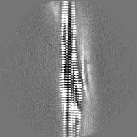
| Images |  emd_52781.png emd_52781.png | 37.9 KB | ||
| Masks | emd_52781_msk_1.map | 30.5 MB | Mask map | |
| Filedesc metadata | emd-52781.cif.gz | 6.1 KB | ||
| Others | emd_52781_half_map_1.map.gzemd_52781_half_map_2.map.gz | 26.9 MB 23.3 MB | ||
| Archive directory |  http://ftp.pdbj.org/pub/emdb/structures/EMD-52781ftp://ftp.pdbj.org/pub/emdb/structures/EMD-52781 http://ftp.pdbj.org/pub/emdb/structures/EMD-52781ftp://ftp.pdbj.org/pub/emdb/structures/EMD-52781 | HTTPS FTP |
-Related structure data
| Related structure data |  9iahMC M: atomic model generated by this map C: citing same article ( |
|---|---|
| Similar structure data |
-Links
| EMDB pages | EMDB (EBI/PDBe) / EMDataResource |
|---|---|
| Related items in Molecule of the Month |


-Map
| File | Download / File: emd_52781.map.gz / Format: CCP4 / Size: 30.5 MB / Type: IMAGE STORED AS FLOATING POINT NUMBER (4 BYTES) | ||||||||||||||||||||||||||||||||||||
|---|---|---|---|---|---|---|---|---|---|---|---|---|---|---|---|---|---|---|---|---|---|---|---|---|---|---|---|---|---|---|---|---|---|---|---|---|---|
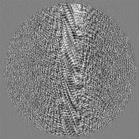
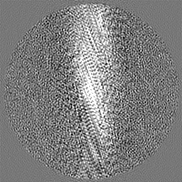
| Projections & slices | Image control
Images are generated by Spider. | ||||||||||||||||||||||||||||||||||||
| Voxel size | X=Y=Z: 1.3 Å | ||||||||||||||||||||||||||||||||||||
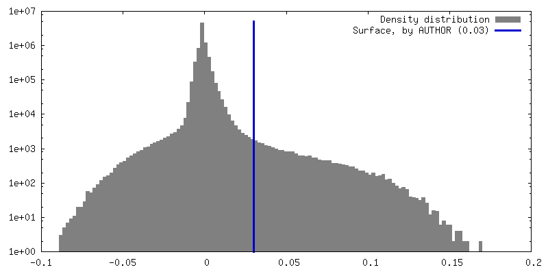
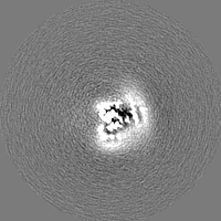
| Density |
| ||||||||||||||||||||||||||||||||||||
| Symmetry | Space group: 1 | ||||||||||||||||||||||||||||||||||||
| Details | EMDB XML:
|
 Z (Sec.)
 Y (Row.)
 X (Col.)


-Supplemental data
-Mask #1
| File | emd_52781_msk_1.map | ||||||||||||
|---|---|---|---|---|---|---|---|---|---|---|---|---|---|
| Projections & Slices |
| ||||||||||||
| Density Histograms |


-Half map: #1
| File | emd_52781_half_map_1.map | ||||||||||||
|---|---|---|---|---|---|---|---|---|---|---|---|---|---|
| Projections & Slices |
| ||||||||||||
| Density Histograms |


-Half map: #2
| File | emd_52781_half_map_2.map | ||||||||||||
|---|---|---|---|---|---|---|---|---|---|---|---|---|---|
| Projections & Slices |
| ||||||||||||
| Density Histograms |


- Sample components
-Entire : Beta-lactoglobulin fibril
| Entire | Name: Beta-lactoglobulin fibril |
|---|---|
| Components |
|
-Supramolecule #1: Beta-lactoglobulin fibril
| Supramolecule | Name: Beta-lactoglobulin fibril / type: complex / ID: 1 / Parent: 0 / Macromolecule list: all |
|---|---|
| Source (natural) | Organism: |
-Macromolecule #1: Beta-lactoglobulin
| Macromolecule | Name: Beta-lactoglobulin / type: protein_or_peptide / ID: 1 / Number of copies: 5 / Enantiomer: LEVO |
|---|---|
| Source (natural) | Organism: |
| Molecular weight | Theoretical: 3.627232 KDa |
| Sequence | String: LIVTQTMKGL DIQKVAGTWY SLAMAASDIS LLDA UniProtKB: Beta-lactoglobulin |
-Experimental details
-Structure determination
| Method | cryo EM |
|---|---|
 Processing Processing | helical reconstruction |
| Aggregation state | filament |
-Sample preparation
| Concentration | 7 mg/mL |
|---|---|
| Buffer | pH: 2.5 / Component - Concentration: 25.0 mM / Component - Name: Citric acid-sodium phosphate |
| Grid | Model: Quantifoil R1.2/1.3 / Material: COPPER / Mesh: 300 / Support film - Material: CARBON / Support film - topology: CONTINUOUS / Support film - Film thickness: 1 / Pretreatment - Type: GLOW DISCHARGE / Pretreatment - Time: 30 sec. / Pretreatment - Atmosphere: AIR / Pretreatment - Pressure: 39.0 kPa Details: PELCO easiGLOW Glow discharge cleaning system using 25 mA for 30 s. |
| Vitrification | Cryogen name: ETHANE-PROPANE / Chamber humidity: 100 % / Chamber temperature: 295.15 K / Instrument: FEI VITROBOT MARK IV Details: 3.7 ul sample was applied and blotted for 6 s after a wait time of 30 s with a force of 0. |
| Details | This sample was monodisperse. The sample was prepared at 7 mg/ml and diluted 15 times for EM grid. |
- Electron microscopy
| Microscope | TFS KRIOS |
|---|---|
| Temperature | Min: 88.15 K / Max: 93.15 K |
| Specialist optics | Energy filter - Name: GIF Bioquantum / Energy filter - Slit width: 20 eV |
| Image recording | Film or detector model: GATAN K3 BIOQUANTUM (6k x 4k) / Detector mode: COUNTING / Digitization - Dimensions - Width: 5760 pixel / Digitization - Dimensions - Height: 4092 pixel / Number grids imaged: 2 / Number real images: 16475 / Average exposure time: 1.0 sec. / Average electron dose: 57.5 e/Å2 |
| Electron beam | Acceleration voltage: 300 kV / Electron source:  FIELD EMISSION GUN FIELD EMISSION GUN |
| Electron optics | C2 aperture diameter: 100.0 µm / Calibrated defocus max: 2.5 µm / Calibrated defocus min: 0.5 µm / Illumination mode: FLOOD BEAM / Imaging mode: BRIGHT FIELD / Cs: 2.7 mm / Nominal defocus max: 2.5 µm / Nominal defocus min: 0.5 µm / Nominal magnification: 130000 |
| Sample stage | Specimen holder model: FEI TITAN KRIOS AUTOGRID HOLDER / Cooling holder cryogen: NITROGEN |
| Experimental equipment |  Model: Titan Krios / Image courtesy: FEI Company |

