 ムービー
ムービー コントローラー


+ データを開く

- 基本情報
基本情報
| 登録情報 | データベース: EMDB / ID: EMD-5269 | |||||||||
|---|---|---|---|---|---|---|---|---|---|---|
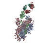
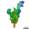
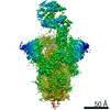
| タイトル | 3D reconstruction of yeast coatomer | |||||||||
 マップデータ マップデータ | 3D reconstruction of yeast coatomer | |||||||||
 試料 試料 |
| |||||||||
 キーワード キーワード | coatomer / COP1 vesicle / vesicular trafficking / coat complex | |||||||||
| 生物種 |  | |||||||||
| 手法 | 単粒子再構成法 / クライオ電子顕微鏡法 / ネガティブ染色法 / 解像度: 36.0 Å | |||||||||
 データ登録者 データ登録者 | Yip CK / Walz T | |||||||||
 引用 引用 | ジャーナル: J Mol Biol / 年: 2011 タイトル: Molecular structure and flexibility of the yeast coatomer as revealed by electron microscopy. 著者: Calvin K Yip / Thomas Walz /  要旨: Coat protein complex I (COPI)-coated vesicles, one of three major types of vesicular carriers in the cell, mediate the early secretory pathway and retrograde transport from the Golgi to the ...Coat protein complex I (COPI)-coated vesicles, one of three major types of vesicular carriers in the cell, mediate the early secretory pathway and retrograde transport from the Golgi to the endoplasmic reticulum. COPI vesicles are generated through activation of the regulatory GTPase Arf1 at the donor membrane and the subsequent recruitment of coatomer, a coat protein complex consisting of seven stably associated components. Coatomer functions in binding and sequestering cargo molecules and assembles into a polymeric protein shell that encompasses the surface of COPI vesicles. Little is known about the structural properties of this heptameric complex. We have isolated native yeast coatomer and examined its structure and subunit organization by single-particle electron microscopy. Our analyses provide the first three-dimensional picture of the complete coatomer and reveal substantial conformational flexibility likely to be critical for its scaffolding function. | |||||||||
| 履歴 |
|
- 構造の表示
構造の表示
| ムービー |
 ムービービューア ムービービューア |
|---|---|
| 構造ビューア | EMマップ: SurfViewMolmilJmol/JSmol |
| 添付画像 |

- ダウンロードとリンク
-EMDBアーカイブ
| マップデータ | emd_5269.map.gz | 2.6 MB | EMDBマップデータ形式 | |
|---|---|---|---|---|
| ヘッダ (付随情報) | emd-5269-v30.xmlemd-5269.xml | 13.8 KB 13.8 KB | 表示 表示 | EMDBヘッダ |
| 画像 | 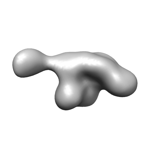 emd_5269_1.png emd_5269_1.png | 25.8 KB | ||
| アーカイブディレクトリ |  http://ftp.pdbj.org/pub/emdb/structures/EMD-5269ftp://ftp.pdbj.org/pub/emdb/structures/EMD-5269 http://ftp.pdbj.org/pub/emdb/structures/EMD-5269ftp://ftp.pdbj.org/pub/emdb/structures/EMD-5269 | HTTPS FTP |
-検証レポート
| 文書・要旨 | emd_5269_validation.pdf.gz | 77.8 KB | 表示 | EMDB検証レポート |
|---|---|---|---|---|
| 文書・詳細版 | emd_5269_full_validation.pdf.gz | 76.9 KB | 表示 | |
| XML形式データ | emd_5269_validation.xml.gz | 494 B | 表示 | |
| アーカイブディレクトリ | https://ftp.pdbj.org/pub/emdb/validation_reports/EMD-5269ftp://ftp.pdbj.org/pub/emdb/validation_reports/EMD-5269 | HTTPS FTP |
-関連構造データ


-リンク
| EMDBのページ | EMDB (EBI/PDBe) / EMDataResource |
|---|
-マップ
| ファイル | ダウンロード / ファイル: emd_5269.map.gz / 形式: CCP4 / 大きさ: 2.7 MB / タイプ: IMAGE STORED AS FLOATING POINT NUMBER (4 BYTES) | ||||||||||||||||||||||||||||||||||||||||||||||||||||||||||||||||||||
|---|---|---|---|---|---|---|---|---|---|---|---|---|---|---|---|---|---|---|---|---|---|---|---|---|---|---|---|---|---|---|---|---|---|---|---|---|---|---|---|---|---|---|---|---|---|---|---|---|---|---|---|---|---|---|---|---|---|---|---|---|---|---|---|---|---|---|---|---|---|
| 注釈 | 3D reconstruction of yeast coatomer | ||||||||||||||||||||||||||||||||||||||||||||||||||||||||||||||||||||
| 投影像・断面図 | 画像のコントロール
画像は Spider により作成 | ||||||||||||||||||||||||||||||||||||||||||||||||||||||||||||||||||||
| ボクセルのサイズ | X=Y=Z: 4.2 Å | ||||||||||||||||||||||||||||||||||||||||||||||||||||||||||||||||||||
| 密度 |
| ||||||||||||||||||||||||||||||||||||||||||||||||||||||||||||||||||||
| 対称性 | 空間群: 1 | ||||||||||||||||||||||||||||||||||||||||||||||||||||||||||||||||||||
| 詳細 | EMDB XML:
CCP4マップ ヘッダ情報:
| ||||||||||||||||||||||||||||||||||||||||||||||||||||||||||||||||||||
 Z (Sec.)
 Y (Row.)
 X (Col.)


-添付データ
- 試料の構成要素
-全体 : Yeast coatomer complex
| 全体 | 名称: Yeast coatomer complex |
|---|---|
| 要素 |
|
-超分子 #1000: Yeast coatomer complex
| 超分子 | 名称: Yeast coatomer complex / タイプ: sample / ID: 1000 / 集合状態: one heteroheptamer / Number unique components: 7 |
|---|---|
| 分子量 | 理論値: 570 KDa |
-分子 #1: Cop1
| 分子 | 名称: Cop1 / タイプ: protein_or_peptide / ID: 1 / Name.synonym: alpha / コピー数: 1 / 組換発現: No / データベース: NCBI |
|---|---|
| 由来(天然) | 生物種: |
| 分子量 | 理論値: 136 KDa |
-分子 #2: Sec26
| 分子 | 名称: Sec26 / タイプ: protein_or_peptide / ID: 2 / Name.synonym: beta / コピー数: 1 / 組換発現: No / データベース: NCBI |
|---|---|
| 由来(天然) | 生物種: |
| 分子量 | 理論値: 109 KDa |
-分子 #3: Sec27
| 分子 | 名称: Sec27 / タイプ: protein_or_peptide / ID: 3 / Name.synonym: beta' / コピー数: 1 / 組換発現: No / データベース: NCBI |
|---|---|
| 由来(天然) | 生物種: |
| 分子量 | 理論値: 109 KDa |
-分子 #4: Sec21
| 分子 | 名称: Sec21 / タイプ: protein_or_peptide / ID: 4 / Name.synonym: gamma / コピー数: 1 / 組換発現: No / データベース: NCBI |
|---|---|
| 由来(天然) | 生物種: |
| 分子量 | 理論値: 105 KDa |
-分子 #5: Ret2
| 分子 | 名称: Ret2 / タイプ: protein_or_peptide / ID: 5 / Name.synonym: delta / コピー数: 1 / 組換発現: No / データベース: NCBI |
|---|---|
| 由来(天然) | 生物種: |
| 分子量 | 理論値: 61 KDa |
-分子 #6: Sec28
| 分子 | 名称: Sec28 / タイプ: protein_or_peptide / ID: 6 / Name.synonym: epsilon / コピー数: 1 / 組換発現: No / データベース: NCBI |
|---|---|
| 由来(天然) | 生物種: |
| 分子量 | 理論値: 34 KDa |
-分子 #7: Ret3
| 分子 | 名称: Ret3 / タイプ: protein_or_peptide / ID: 7 / Name.synonym: zeta / コピー数: 1 / 組換発現: No / データベース: NCBI |
|---|---|
| 由来(天然) | 生物種: |
| 分子量 | 理論値: 22 KDa |
-実験情報
-構造解析
| 手法 | ネガティブ染色法, クライオ電子顕微鏡法 |
|---|---|
 解析 解析 | 単粒子再構成法 |
| 試料の集合状態 | particle |
-試料調製
| 濃度 | 0.02 mg/mL |
|---|---|
| 緩衝液 | pH: 8 詳細: 20 mM Tris, pH 8.0, 150 mM NaCl, 10% glycerol, 0.005% NP40, 0.5 mM DTT |
| 染色 | タイプ: NEGATIVE 詳細: Protein solution was adsorbed onto grids for 30 seconds before staining with 0.75% uranyl formate |
| 凍結 | 凍結剤: NITROGEN / チャンバー内温度: 100 K / 装置: OTHER / 手法: Manual plunging into liquid nitrogen |
- 電子顕微鏡法
| 顕微鏡 | FEI TECNAI F20 |
|---|---|
| 温度 | 平均: 100 K |
| 詳細 | Low dose imaging, same specimen area imaged twice |
| 日付 | 2009年3月1日 |
| 撮影 | カテゴリ: FILM / フィルム・検出器のモデル: KODAK SO-163 FILM / デジタル化 - スキャナー: ZEISS SCAI / デジタル化 - サンプリング間隔: 21 µm |
| Tilt angle min | 0 |
| 電子線 | 加速電圧: 200 kV / 電子線源:  FIELD EMISSION GUN FIELD EMISSION GUN |
| 電子光学系 | 倍率(補正後): 51159 / 照射モード: FLOOD BEAM / 撮影モード: BRIGHT FIELD / Cs: 1.4 mm / 最大 デフォーカス(公称値): 2.5 µm / 最小 デフォーカス(公称値): 1.8 µm / 倍率(公称値): 50000 |
| 試料ステージ | 試料ホルダー: Side entry liquid nitrogen-cooled cryo specimen holder 試料ホルダーモデル: OTHER / Tilt angle max: 50 |
| 実験機器 |  モデル: Tecnai F20 / 画像提供: FEI Company |
-画像解析
| 最終 再構成 | アルゴリズム: OTHER / 解像度のタイプ: BY AUTHOR / 解像度: 36.0 Å / 解像度の算出法: FSC 0.5 CUT-OFF / ソフトウェア - 名称: SPIDER / 詳細: Final map is filtered to 36 Angstroms resolution |
|---|---|
| 最終 角度割当 | 詳細: SPIDER:theta 45 degrees, phi 45 degrees |
