 ムービー
ムービー コントローラー


+ データを開く

- 基本情報
基本情報
| 登録情報 | データベース: EMDB / ID: EMD-5242 | |||||||||
|---|---|---|---|---|---|---|---|---|---|---|
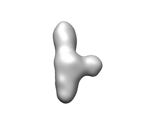
| タイトル | B. subtilis RNase P RNA Specificity domain folding intermediate | |||||||||
 マップデータ マップデータ | This is a map of the folding intermediate of B. subtilis RNase P Specificity domain | |||||||||
 試料 試料 |
| |||||||||
 キーワード キーワード | RNA folding intermediate RNase P Specificity domain | |||||||||
| 生物種 |  | |||||||||
| 手法 | 単粒子再構成法 / クライオ電子顕微鏡法 / 解像度: 15.2 Å | |||||||||
 データ登録者 データ登録者 | Baird NJ / Ludtke SJ / Khant H / Chiu W / Pan T / Sosnick TR | |||||||||
 引用 引用 | ジャーナル: J Am Chem Soc / 年: 2010 タイトル: Discrete structure of an RNA folding intermediate revealed by cryo-electron microscopy. 著者: Nathan J Baird / Steven J Ludtke / Htet Khant / Wah Chiu / Tao Pan / Tobin R Sosnick /  要旨: RNA folding occurs via a series of transitions between metastable intermediate states. It is unknown whether folding intermediates are discrete structures folding along defined pathways or ...RNA folding occurs via a series of transitions between metastable intermediate states. It is unknown whether folding intermediates are discrete structures folding along defined pathways or heterogeneous ensembles folding along broad landscapes. We use cryo-electron microscopy and single-particle image reconstruction to determine the structure of the major folding intermediate of the specificity domain of a ribonuclease P ribozyme. Our results support the existence of a discrete conformation for this folding intermediate. | |||||||||
| 履歴 |
|
- 構造の表示
構造の表示
| ムービー |
 ムービービューア ムービービューア |
|---|---|
| 構造ビューア | EMマップ: SurfViewMolmilJmol/JSmol |
| 添付画像 |
- ダウンロードとリンク
-EMDBアーカイブ
| マップデータ | emd_5242.map.gz | 14.1 MB | EMDBマップデータ形式 | |
|---|---|---|---|---|
| ヘッダ (付随情報) | emd-5242-v30.xmlemd-5242.xml | 10.1 KB 10.1 KB | 表示 表示 | EMDBヘッダ |
| 画像 | emd_5242_1.tif | 34.7 KB | ||
| アーカイブディレクトリ |  http://ftp.pdbj.org/pub/emdb/structures/EMD-5242ftp://ftp.pdbj.org/pub/emdb/structures/EMD-5242 http://ftp.pdbj.org/pub/emdb/structures/EMD-5242ftp://ftp.pdbj.org/pub/emdb/structures/EMD-5242 | HTTPS FTP |
-関連構造データ


-リンク
| EMDBのページ | EMDB (EBI/PDBe) / EMDataResource |
|---|
-マップ
| ファイル | ダウンロード / ファイル: emd_5242.map.gz / 形式: CCP4 / 大きさ: 15.3 MB / タイプ: IMAGE STORED AS FLOATING POINT NUMBER (4 BYTES) | ||||||||||||||||||||||||||||||||||||||||||||||||||||||||||||||||||||
|---|---|---|---|---|---|---|---|---|---|---|---|---|---|---|---|---|---|---|---|---|---|---|---|---|---|---|---|---|---|---|---|---|---|---|---|---|---|---|---|---|---|---|---|---|---|---|---|---|---|---|---|---|---|---|---|---|---|---|---|---|---|---|---|---|---|---|---|---|---|
| 注釈 | This is a map of the folding intermediate of B. subtilis RNase P Specificity domain | ||||||||||||||||||||||||||||||||||||||||||||||||||||||||||||||||||||
| 投影像・断面図 | 画像のコントロール
画像は Spider により作成 | ||||||||||||||||||||||||||||||||||||||||||||||||||||||||||||||||||||
| ボクセルのサイズ | X=Y=Z: 1.81 Å | ||||||||||||||||||||||||||||||||||||||||||||||||||||||||||||||||||||
| 密度 |
| ||||||||||||||||||||||||||||||||||||||||||||||||||||||||||||||||||||
| 対称性 | 空間群: 1 | ||||||||||||||||||||||||||||||||||||||||||||||||||||||||||||||||||||
| 詳細 | EMDB XML:
CCP4マップ ヘッダ情報:
| ||||||||||||||||||||||||||||||||||||||||||||||||||||||||||||||||||||
 Z (Sec.)
 Y (Row.)
 X (Col.)


-添付データ
- 試料の構成要素
-全体 : B. subtilis RNase P RNA Specificity domain folding intermediate
| 全体 | 名称: B. subtilis RNase P RNA Specificity domain folding intermediate |
|---|---|
| 要素 |
|
-超分子 #1000: B. subtilis RNase P RNA Specificity domain folding intermediate
| 超分子 | 名称: B. subtilis RNase P RNA Specificity domain folding intermediate タイプ: sample / ID: 1000 / 詳細: none / 集合状態: Monomer of Specificity domain / Number unique components: 1 |
|---|---|
| 分子量 | 理論値: 50 KDa / 手法: Calculation from nucleotide sequence, 154mer RNA |
-分子 #1: RNA
| 分子 | 名称: RNA / タイプ: rna / ID: 1 / Name.synonym: RNase P RNA Specificity domain / 分類: OTHER / Structure: OTHER / Synthetic?: No |
|---|---|
| 由来(天然) | 生物種: |
| 分子量 | 理論値: 50 KDa |
| 配列 | 文字列: GCGAGCCUAG CGAAGUCAUA AGCUAGGGCA GUCUUUAGAG GCUGACGGCA GGAAAAAAGC CUACGUCUUC GGAUAUGGCU GAGUAUCCUU GAAAGUGCCA CAGUGACGAA GUCUCACUAG AAAUGGUGAG AGUGGAACGC GGUAAACCCC UCGC |
-実験情報
-構造解析
| 手法 | クライオ電子顕微鏡法 |
|---|---|
 解析 解析 | 単粒子再構成法 |
| 試料の集合状態 | particle |
-試料調製
| 濃度 | 1 mg/mL |
|---|---|
| 緩衝液 | pH: 8 / 詳細: 1 mM MgCl2, 20 mM TrisHCl pH 8 |
| グリッド | 詳細: 400 mesh carbon grid |
| 凍結 | 凍結剤: ETHANE / チャンバー内湿度: 100 % / チャンバー内温度: 77 K / 装置: FEI VITROBOT MARK III / 詳細: Vitrification instrument: FEI Vitrobot mark III / 手法: 2 blots 1 second each before plunging |
- 電子顕微鏡法
| 顕微鏡 | JEOL 2010F |
|---|---|
| 温度 | 最低: 93 K / 最高: 95 K / 平均: 94.1 K |
| アライメント法 | Legacy - 非点収差: object astigmatism correction made at 400,000 times magnification |
| 日付 | 2006年3月10日 |
| 撮影 | カテゴリ: CCD フィルム・検出器のモデル: GATAN ULTRASCAN 4000 (4k x 4k) 実像数: 100 / 平均電子線量: 16 e/Å2 |
| 電子線 | 加速電圧: 200 kV / 電子線源:  FIELD EMISSION GUN FIELD EMISSION GUN |
| 電子光学系 | 照射モード: FLOOD BEAM / 撮影モード: BRIGHT FIELD / Cs: 2 mm / 最大 デフォーカス(公称値): 3.5 µm / 最小 デフォーカス(公称値): 1.0 µm / 倍率(公称値): 60000 |
| 試料ステージ | 試料ホルダー: single tilt cryo-holder / 試料ホルダーモデル: GATAN LIQUID NITROGEN |
-画像解析
| 詳細 | The particles were selected using an automatic selection program and then inspected manually |
|---|---|
| 最終 再構成 | アルゴリズム: OTHER / 解像度のタイプ: BY AUTHOR / 解像度: 15.2 Å / 解像度の算出法: FSC 0.5 CUT-OFF / ソフトウェア - 名称: EMAN 詳細: FSC gives a resolution of 15.2 A, but the model was low-pass filtered to 26 A, corresponding to the first zero-crossing of the data. CTF correction was not performed. 使用した粒子像数: 11600 |
| 最終 2次元分類 | クラス数: 60 |
