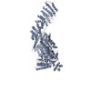
Movie
Movie Controller

[English] 日本語
 Yorodumi
- EMDB-52295: Consensus map of human separase bound to SCC1 (310-550 aa) and SA2 -
+ Open data

- Basic information
| Entry |  | |||||||||
|---|---|---|---|---|---|---|---|---|---|---|
| Title | Consensus map of human separase bound to SCC1 (310-550 aa) and SA2 | |||||||||
 Map data Map data | Consensus map (unsharpened) of human separase bound to SCC1 (310-550 aa) and SA2 | |||||||||
 Sample Sample |
| |||||||||
 Keywords Keywords | Separase / cell cycle / SCC1 / RAD21 / protease / chromosome segregation / Auto-cleavage / SA1/2 / cohesin | |||||||||
| Biological species |  Homo sapiens (human) Homo sapiens (human) | |||||||||
| Method | single particle reconstruction / cryo EM / Resolution: 3.4 Å | |||||||||
 Authors Authors | Yu J / Schmidt S / Botto M / Boland A | |||||||||
| Funding support |  Switzerland, 2 items Switzerland, 2 items
| |||||||||
 Citation Citation | Journal: Sci Adv / Year: 2025 Title: Substrate recognition by human separase. Authors: Jun Yu / Sophia Schmidt / Margherita Botto / Kitaik Lee / Chloe M Ghent / Jonah M Goodfried / Andrew Howe / Francis J O'Reilly / David O Morgan / Andreas Boland /  Abstract: The cohesin complex encircles sister chromatids in early mitosis. At anaphase onset, sister separation is triggered by the proteolytic cleavage of the cohesin subunit SCC1/RAD21 by separase. SCC1 ...The cohesin complex encircles sister chromatids in early mitosis. At anaphase onset, sister separation is triggered by the proteolytic cleavage of the cohesin subunit SCC1/RAD21 by separase. SCC1 contains two cleavage sites, where cleavage is stimulated by SCC1 phosphorylation. Substrate recognition and cleavage are only partly understood. Here, we determined structures of human separase in apo- or substrate-bound forms that, together with biochemical analysis, provide critical insights into separase cleavage regulation. We verify the first SCC1 cleavage site and reassign the second. We show that substrates, including separase autocleavage sites and the two SCC1 cleavage sites, interact with docking sites in separase, including five phosphate-binding sites. We also describe the interaction between the cohesin subunit SA1/SA2 and separase, which promotes cleavage at the second SCC1 site. Using cross-linking mass spectrometry and cryo-electron microscopy, we propose how cohesin is targeted by human separase. Our work provides an extensive functional and structural framework that explains a key event in cell division. | |||||||||
| History |
|
- Structure visualization
| Supplemental images |
|---|

- Downloads & links
-EMDB archive
| Map data | emd_52295.map.gz | 256.5 MB |  EMDB map data format EMDB map data format | |
|---|---|---|---|---|
| Header (meta data) | emd-52295-v30.xmlemd-52295.xml | 22.3 KB 22.3 KB | Display Display | EMDB header |
| Images |  emd_52295.png emd_52295.png | 50.4 KB | ||
| Filedesc metadata | emd-52295.cif.gz | 5.2 KB | ||
| Others | emd_52295_half_map_1.map.gzemd_52295_half_map_2.map.gz | 475.7 MB 475.7 MB | ||
| Archive directory |  http://ftp.pdbj.org/pub/emdb/structures/EMD-52295ftp://ftp.pdbj.org/pub/emdb/structures/EMD-52295 http://ftp.pdbj.org/pub/emdb/structures/EMD-52295ftp://ftp.pdbj.org/pub/emdb/structures/EMD-52295 | HTTPS FTP |
-Related structure data


-Links
| EMDB pages | EMDB (EBI/PDBe) / EMDataResource |
|---|
-Map
| File | Download / File: emd_52295.map.gz / Format: CCP4 / Size: 512 MB / Type: IMAGE STORED AS FLOATING POINT NUMBER (4 BYTES) | ||||||||||||||||||||||||||||||||||||
|---|---|---|---|---|---|---|---|---|---|---|---|---|---|---|---|---|---|---|---|---|---|---|---|---|---|---|---|---|---|---|---|---|---|---|---|---|---|
| Annotation | Consensus map (unsharpened) of human separase bound to SCC1 (310-550 aa) and SA2 | ||||||||||||||||||||||||||||||||||||
| Projections & slices | Image control
Images are generated by Spider. | ||||||||||||||||||||||||||||||||||||
| Voxel size | X=Y=Z: 0.9024 Å | ||||||||||||||||||||||||||||||||||||
| Density |
| ||||||||||||||||||||||||||||||||||||
| Symmetry | Space group: 1 | ||||||||||||||||||||||||||||||||||||
| Details | EMDB XML:
|
 Z (Sec.)
 Y (Row.)
 X (Col.)


-Supplemental data
-Half map: Consensus half map A of human separase bound...
| File | emd_52295_half_map_1.map | ||||||||||||
|---|---|---|---|---|---|---|---|---|---|---|---|---|---|
| Annotation | Consensus half map A of human separase bound to SCC1 (310-550 aa) and SA2 | ||||||||||||
| Projections & Slices |
| ||||||||||||
| Density Histograms |


-Half map: Consensus half map B of human separase bound...
| File | emd_52295_half_map_2.map | ||||||||||||
|---|---|---|---|---|---|---|---|---|---|---|---|---|---|
| Annotation | Consensus half map B of human separase bound to SCC1 (310-550 aa) and SA2 | ||||||||||||
| Projections & Slices |
| ||||||||||||
| Density Histograms |


- Sample components
-Entire : Human separase bound to SCC1 (310-550 aa) and SA2
| Entire | Name: Human separase bound to SCC1 (310-550 aa) and SA2 |
|---|---|
| Components |
|
-Supramolecule #1: Human separase bound to SCC1 (310-550 aa) and SA2
| Supramolecule | Name: Human separase bound to SCC1 (310-550 aa) and SA2 / type: complex / ID: 1 / Parent: 0 / Macromolecule list: #1-#2 |
|---|---|
| Source (natural) | Organism: Homo sapiens (human) |
| Molecular weight | Theoretical: 400 KDa |
-Experimental details
-Structure determination
| Method | cryo EM |
|---|---|
 Processing Processing | single particle reconstruction |
| Aggregation state | particle |
-Sample preparation
| Concentration | 0.1 mg/mL | ||||||||||||
|---|---|---|---|---|---|---|---|---|---|---|---|---|---|
| Buffer | pH: 8 Component:
| ||||||||||||
| Grid | Model: Quantifoil R1.2/1.3 / Material: GOLD / Mesh: 300 / Support film - Material: GRAPHENE OXIDE / Support film - topology: CONTINUOUS / Support film - Film thickness: 1 | ||||||||||||
| Vitrification | Cryogen name: ETHANE / Chamber humidity: 90 % / Chamber temperature: 277.15 K / Instrument: LEICA EM GP |
- Electron microscopy
| Microscope | FEI TALOS ARCTICA |
|---|---|
| Specialist optics | Energy filter - Name: TFS Selectris X / Energy filter - Slit width: 10 eV |
| Image recording | Film or detector model: TFS FALCON 4i (4k x 4k) / Detector mode: COUNTING / Digitization - Dimensions - Width: 4096 pixel / Digitization - Dimensions - Height: 4096 pixel / Number grids imaged: 16 / Number real images: 57012 / Average electron dose: 50.0 e/Å2 |
| Electron beam | Acceleration voltage: 200 kV / Electron source:  FIELD EMISSION GUN FIELD EMISSION GUN |
| Electron optics | C2 aperture diameter: 50.0 µm / Illumination mode: FLOOD BEAM / Imaging mode: BRIGHT FIELD / Cs: 2.7 mm / Nominal defocus max: 2.0 µm / Nominal defocus min: 0.6 µm / Nominal magnification: 130000 |
| Sample stage | Specimen holder model: FEI TITAN KRIOS AUTOGRID HOLDER / Cooling holder cryogen: NITROGEN |
| Experimental equipment | 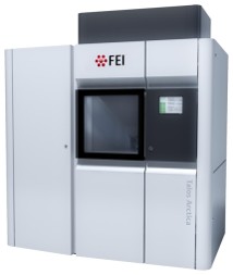 Model: Talos Arctica / Image courtesy: FEI Company |