Movie
Movie Controller


+ Open data

- Basic information
| Entry |  | |||||||||
|---|---|---|---|---|---|---|---|---|---|---|
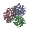
| Title | Transient activated state of BetP-M150E | |||||||||
 Map data Map data | ||||||||||
 Sample Sample |
| |||||||||
 Keywords Keywords | Glycine betaine transporter BetP / TRANSPORT PROTEIN | |||||||||
| Function / homology | BCCT transporter, conserved site / BCCT family of transporters signature. / BCCT transporter family / BCCT, betaine/carnitine/choline family transporter / symporter activity / metal ion binding / identical protein binding / plasma membrane / Glycine betaine transporter BetP Function and homology information Function and homology information | |||||||||
| Biological species |  Corynebacterium glutamicum (bacteria) Corynebacterium glutamicum (bacteria) | |||||||||
| Method | single particle reconstruction / cryo EM / Resolution: 2.74 Å | |||||||||
 Authors Authors | Urbansky K / Gauthier-Manuel L / Fu L / Madej MG / Ziegler C | |||||||||
| Funding support | 1 items
| |||||||||
 Citation Citation | Journal: To be published Title: Structural evidence for a link between substrate promiscuity and transport regulation in BetP mutants Authors: Urbansky K / Fu L / Klinger S / Madej MG / Ziegler C | |||||||||
| History |
|
- Structure visualization
| Supplemental images |
|---|

- Downloads & links
-EMDB archive
| Map data | emd_52232.map.gz | 118 MB | EMDB map data format | |
|---|---|---|---|---|
| Header (meta data) | emd-52232-v30.xmlemd-52232.xml | 17.7 KB 17.7 KB | Display Display | EMDB header |
| FSC (resolution estimation) | emd_52232_fsc.xml | 10.6 KB | Display | FSC data file |
| Images |  emd_52232.png emd_52232.png | 125.3 KB | ||
| Filedesc metadata | emd-52232.cif.gz | 6.2 KB | ||
| Others | emd_52232_half_map_1.map.gzemd_52232_half_map_2.map.gz | 116.2 MB 116.2 MB | ||
| Archive directory |  http://ftp.pdbj.org/pub/emdb/structures/EMD-52232ftp://ftp.pdbj.org/pub/emdb/structures/EMD-52232 http://ftp.pdbj.org/pub/emdb/structures/EMD-52232ftp://ftp.pdbj.org/pub/emdb/structures/EMD-52232 | HTTPS FTP |
-Related structure data
| Related structure data |  9hkmMC  9hklC M: atomic model generated by this map C: citing same article ( |
|---|---|
| Similar structure data |

-Links
| EMDB pages | EMDB (EBI/PDBe) / EMDataResource |
|---|
-Map
| File | Download / File: emd_52232.map.gz / Format: CCP4 / Size: 125 MB / Type: IMAGE STORED AS FLOATING POINT NUMBER (4 BYTES) | ||||||||||||||||||||||||||||||||||||
|---|---|---|---|---|---|---|---|---|---|---|---|---|---|---|---|---|---|---|---|---|---|---|---|---|---|---|---|---|---|---|---|---|---|---|---|---|---|
| Projections & slices | Image control
Images are generated by Spider. | ||||||||||||||||||||||||||||||||||||
| Voxel size | X=Y=Z: 0.7891 Å | ||||||||||||||||||||||||||||||||||||
| Density |
| ||||||||||||||||||||||||||||||||||||
| Symmetry | Space group: 1 | ||||||||||||||||||||||||||||||||||||
| Details | EMDB XML:
|
 Z (Sec.)
 Y (Row.)
 X (Col.)


-Supplemental data
-Half map: #2
| File | emd_52232_half_map_1.map | ||||||||||||
|---|---|---|---|---|---|---|---|---|---|---|---|---|---|
| Projections & Slices |
| ||||||||||||
| Density Histograms |


-Half map: #1
| File | emd_52232_half_map_2.map | ||||||||||||
|---|---|---|---|---|---|---|---|---|---|---|---|---|---|
| Projections & Slices |
| ||||||||||||
| Density Histograms |


- Sample components
-Entire : Glycine betaine transporter BetP-M150E
| Entire | Name: Glycine betaine transporter BetP-M150E |
|---|---|
| Components |
|
-Supramolecule #1: Glycine betaine transporter BetP-M150E
| Supramolecule | Name: Glycine betaine transporter BetP-M150E / type: complex / ID: 1 / Parent: 0 / Macromolecule list: #1 |
|---|---|
| Source (natural) | Organism: Corynebacterium glutamicum (bacteria) |
-Macromolecule #1: Glycine betaine transporter BetP
| Macromolecule | Name: Glycine betaine transporter BetP / type: protein_or_peptide / ID: 1 / Number of copies: 3 / Enantiomer: LEVO |
|---|---|
| Source (natural) | Organism: Corynebacterium glutamicum (bacteria) |
| Molecular weight | Theoretical: 65.296008 KDa |
| Recombinant expression | Organism: |
| Sequence | String: WSHPQFEKMT TSDPNPKPIV EDAQPEQITA TEELAGLLEN PTNLEGKLAD AEEEIILEGE DTQASLNWSV IVPALVIVLA TVVWGIGFK DSFTNFASSA LSAVVDNLGW AFILFGTVFV FFIVVIAASK FGTIRLGRID EAPEFRTVSW ISMMFAAGEG I GLMFYGTT ...String: WSHPQFEKMT TSDPNPKPIV EDAQPEQITA TEELAGLLEN PTNLEGKLAD AEEEIILEGE DTQASLNWSV IVPALVIVLA TVVWGIGFK DSFTNFASSA LSAVVDNLGW AFILFGTVFV FFIVVIAASK FGTIRLGRID EAPEFRTVSW ISMMFAAGEG I GLMFYGTT EPLTFYRNGV PGHDEHNVGV AMSTTMFHWT LHPWAIYAIV GLAIAYSTFR VGRKQLLSSA FVPLIGEKGA EG WLGKLID ILAIIATVFG TACSLGLGAL QIGAGLSAAN IIEDPSDWTI VGIVSVLTLA FIFSAISGVG KGIQYLSNAN MVL AALLAI FVFVVGPTVS ILNLLPGSIG NYLSNFFQMA GRTAMSADGT AGEWLGSWTI FYWAWWISWS PFVGMFLARI SRGR SIREF ILGVLLVPAG VSTVWFSIFG GTAIVFEQNG ESIWGDGAAE EQLFGLLHAL PGGQIMGIIA MILLGTFFIT SADSA STVM GTMSQHGQLE ANKWVTAAWG VATAAIGLTL LLSGGDNALS NLQNVTIVAA TPFLFVVIGL MFALVKDLSN DVIYLE YRE QQRFNARLAR ERRVHNEHRK RELAAKRRRE RKASGAGKRR UniProtKB: Glycine betaine transporter BetP |
-Macromolecule #2: (1S)-2-{[{[(2R)-2,3-DIHYDROXYPROPYL]OXY}(HYDROXY)PHOSPHORYL]OXY}-...
| Macromolecule | Name: (1S)-2-{[{[(2R)-2,3-DIHYDROXYPROPYL]OXY}(HYDROXY)PHOSPHORYL]OXY}-1-[(PALMITOYLOXY)METHYL]ETHYL STEARATE type: ligand / ID: 2 / Number of copies: 3 / Formula: PGT |
|---|---|
| Molecular weight | Theoretical: 751.023 Da |
| Chemical component information |  ChemComp-PGT: |
-Macromolecule #3: GAMMA-AMINO-BUTANOIC ACID
| Macromolecule | Name: GAMMA-AMINO-BUTANOIC ACID / type: ligand / ID: 3 / Number of copies: 1 / Formula: ABU |
|---|---|
| Molecular weight | Theoretical: 103.12 Da |
| Chemical component information |  ChemComp-ABU: |
-Macromolecule #4: DODECYL-BETA-D-MALTOSIDE
| Macromolecule | Name: DODECYL-BETA-D-MALTOSIDE / type: ligand / ID: 4 / Number of copies: 1 / Formula: LMT |
|---|---|
| Molecular weight | Theoretical: 510.615 Da |
| Chemical component information |  ChemComp-LMT: |
-Experimental details
-Structure determination
| Method | cryo EM |
|---|---|
 Processing Processing | single particle reconstruction |
| Aggregation state | particle |
-Sample preparation
| Buffer | pH: 7.5 Component:
| ||||||||
|---|---|---|---|---|---|---|---|---|---|
| Vitrification | Cryogen name: ETHANE / Instrument: FEI VITROBOT MARK III |
- Electron microscopy
| Microscope | JEOL CRYO ARM 200 |
|---|---|
| Image recording | Film or detector model: GATAN K2 SUMMIT (4k x 4k) / Detector mode: COUNTING / Average electron dose: 52.66 e/Å2 |
| Electron beam | Acceleration voltage: 200 kV / Electron source:  FIELD EMISSION GUN FIELD EMISSION GUN |
| Electron optics | Illumination mode: FLOOD BEAM / Imaging mode: BRIGHT FIELD / Nominal defocus max: 1.4000000000000001 µm / Nominal defocus min: 0.6 µm |
| Sample stage | Specimen holder model: JEOL CRYOSPECPORTER / Cooling holder cryogen: NITROGEN |