[English] 日本語
 Yorodumi
- EMDB-51768: Cryo-EM structure of taxol-microtubules in complex with the C1 do... -
+ Open data
- Basic information
| Entry |  | |||||||||
|---|---|---|---|---|---|---|---|---|---|---|
| Title | Cryo-EM structure of taxol-microtubules in complex with the C1 domain of GEFH1 | |||||||||
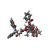
 Map data Map data | Cryo-EM reconstruction of the C1 domain of GEFH1 in complex with taxol-stabilized 13 protofilament microtubule | |||||||||
 Sample Sample |
| |||||||||
 Keywords Keywords | RhoGEF / C1 / Cytoskeleton / Complex / SIGNALING PROTEIN | |||||||||
| Function / homology |  Function and homology information Function and homology informationasymmetric neuroblast division / negative regulation of intrinsic apoptotic signaling pathway in response to osmotic stress / cellular response to muramyl dipeptide / positive regulation of neuron migration / negative regulation of microtubule depolymerization / regulation of Rho protein signal transduction / negative regulation of necroptotic process / cellular hyperosmotic response / positive regulation of axon guidance / regulation of small GTPase mediated signal transduction ...asymmetric neuroblast division / negative regulation of intrinsic apoptotic signaling pathway in response to osmotic stress / cellular response to muramyl dipeptide / positive regulation of neuron migration / negative regulation of microtubule depolymerization / regulation of Rho protein signal transduction / negative regulation of necroptotic process / cellular hyperosmotic response / positive regulation of axon guidance / regulation of small GTPase mediated signal transduction / positive regulation of peptidyl-tyrosine phosphorylation / RHOB GTPase cycle / NRAGE signals death through JNK / RHOA GTPase cycle / bicellular tight junction / microtubule-based process / negative regulation of extrinsic apoptotic signaling pathway via death domain receptors / cytoplasmic microtubule / positive regulation of neuron differentiation / cellular response to interleukin-4 / guanyl-nucleotide exchange factor activity / actin filament organization / intracellular protein transport / : / structural constituent of cytoskeleton / positive regulation of interleukin-6 production / small GTPase binding / microtubule cytoskeleton organization / spindle / ruffle membrane / neuron migration / cell morphogenesis / positive regulation of tumor necrosis factor production / cellular response to tumor necrosis factor / mitotic cell cycle / G alpha (12/13) signalling events / regulation of cell population proliferation / double-stranded RNA binding / microtubule cytoskeleton / cytoplasmic vesicle / microtubule binding / vesicle / Hydrolases; Acting on acid anhydrides; Acting on GTP to facilitate cellular and subcellular movement / microtubule / cytoskeleton / cilium / protein heterodimerization activity / innate immune response / focal adhesion / GTPase activity / ubiquitin protein ligase binding / GTP binding / Golgi apparatus / positive regulation of transcription by RNA polymerase II / protein-containing complex / zinc ion binding / metal ion binding / cytoplasm / cytosol Similarity search - Function | |||||||||
| Biological species |  Homo sapiens (human) / Homo sapiens (human) /  | |||||||||
| Method | single particle reconstruction / cryo EM / Resolution: 3.4 Å | |||||||||
 Authors Authors | Choi SR / Blum T / Steinmetz MO | |||||||||
| Funding support |  Switzerland, 1 items Switzerland, 1 items
| |||||||||
 Citation Citation |  Journal: Cell / Year: 2026 Journal: Cell / Year: 2026Title: Structural basis of microtubule-mediated signal transduction. Authors: Sung Ryul Choi / Thorsten B Blum / Matteo Giono / Bibhas Roy / Ioannis Vakonakis / Dominic Schmid / Nicole Oelgarth / Apisha Ranganathan / Alvar D Gossert / G V Shivashankar / Alfred ...Authors: Sung Ryul Choi / Thorsten B Blum / Matteo Giono / Bibhas Roy / Ioannis Vakonakis / Dominic Schmid / Nicole Oelgarth / Apisha Ranganathan / Alvar D Gossert / G V Shivashankar / Alfred Zippelius / Michel O Steinmetz /    Abstract: Microtubules have long been recognized as upstream mediators of intracellular signaling, but the mechanisms underlying this fundamental function remain elusive. Here, we identify the structural basis ...Microtubules have long been recognized as upstream mediators of intracellular signaling, but the mechanisms underlying this fundamental function remain elusive. Here, we identify the structural basis by which microtubules regulate the guanine nucleotide exchange factor H1 (GEFH1), a key activator of the Ras homolog family member A (RhoA) pathway. We show that specific features of the microtubule lattice bind the C1 domain of GEFH1, leading to the sequestration and inactivation of this signaling protein. Targeted mutations in C1 residues disrupt this interaction, triggering GEFH1 release and activation of RhoA-dependent immune responses. Building on this sequestration-and-release mechanism, we identify microtubule-binding C1 domains in additional signaling proteins, including other guanine nucleotide exchange factors (GEFs), kinases, a GTPase-activating protein (GAP), and a tumor suppressor, and show that microtubule-mediated regulation via C1 domains is conserved in the Ras association domain-containing protein 1A (RASSF1A). Our findings establish a structural framework for understanding how microtubules can function as spatiotemporal signal sensors, integrating and processing diverse signaling pathways to control important cellular processes. | |||||||||
| History |
|
- Structure visualization
| Supplemental images |
|---|
- Downloads & links
-EMDB archive
| Map data |  emd_51768.map.gz emd_51768.map.gz | 14.4 MB |  EMDB map data format EMDB map data format | |
|---|---|---|---|---|
| Header (meta data) |  emd-51768-v30.xml emd-51768-v30.xml emd-51768.xml emd-51768.xml | 27.8 KB 27.8 KB | Display Display |  EMDB header EMDB header |
| Images |  emd_51768.png emd_51768.png | 58.5 KB | ||
| Filedesc metadata |  emd-51768.cif.gz emd-51768.cif.gz | 7.5 KB | ||
| Others |  emd_51768_half_map_1.map.gz emd_51768_half_map_1.map.gz emd_51768_half_map_2.map.gz emd_51768_half_map_2.map.gz | 11.9 MB 11.9 MB | ||
| Archive directory |  http://ftp.pdbj.org/pub/emdb/structures/EMD-51768 http://ftp.pdbj.org/pub/emdb/structures/EMD-51768 ftp://ftp.pdbj.org/pub/emdb/structures/EMD-51768 ftp://ftp.pdbj.org/pub/emdb/structures/EMD-51768 | HTTPS FTP |
-Related structure data
| Related structure data |  9h1oMC  9h75C M: atomic model generated by this map C: citing same article ( |
|---|---|
| Similar structure data | Similarity search - Function & homology  F&H Search F&H Search |
- Links
Links
| EMDB pages |  EMDB (EBI/PDBe) / EMDB (EBI/PDBe) /  EMDataResource EMDataResource |
|---|---|
| Related items in Molecule of the Month |
- Map
Map
| File |  Download / File: emd_51768.map.gz / Format: CCP4 / Size: 15.6 MB / Type: IMAGE STORED AS FLOATING POINT NUMBER (4 BYTES) Download / File: emd_51768.map.gz / Format: CCP4 / Size: 15.6 MB / Type: IMAGE STORED AS FLOATING POINT NUMBER (4 BYTES) | ||||||||||||||||||||||||||||||||||||
|---|---|---|---|---|---|---|---|---|---|---|---|---|---|---|---|---|---|---|---|---|---|---|---|---|---|---|---|---|---|---|---|---|---|---|---|---|---|
| Annotation | Cryo-EM reconstruction of the C1 domain of GEFH1 in complex with taxol-stabilized 13 protofilament microtubule | ||||||||||||||||||||||||||||||||||||
| Projections & slices | Image control
Images are generated by Spider. | ||||||||||||||||||||||||||||||||||||
| Voxel size | X=Y=Z: 1.058 Å | ||||||||||||||||||||||||||||||||||||
| Density |
| ||||||||||||||||||||||||||||||||||||
| Symmetry | Space group: 1 | ||||||||||||||||||||||||||||||||||||
| Details | EMDB XML:
|
-Supplemental data
-Half map: Half map 1: Cryo-EM reconstruction of the C1...
| File | emd_51768_half_map_1.map | ||||||||||||
|---|---|---|---|---|---|---|---|---|---|---|---|---|---|
| Annotation | Half map 1: Cryo-EM reconstruction of the C1 domain of GEFH1 in complex with taxol-stabilized 13 protofilament microtubule | ||||||||||||
| Projections & Slices |
| ||||||||||||
| Density Histograms |
-Half map: Half map 2: Cryo-EM reconstruction of the C1...
| File | emd_51768_half_map_2.map | ||||||||||||
|---|---|---|---|---|---|---|---|---|---|---|---|---|---|
| Annotation | Half map 2: Cryo-EM reconstruction of the C1 domain of GEFH1 in complex with taxol-stabilized 13 protofilament microtubule | ||||||||||||
| Projections & Slices |
| ||||||||||||
| Density Histograms |
- Sample components
+Entire : The C1 domain of GEFH1 in complex with microtubule
+Supramolecule #1: The C1 domain of GEFH1 in complex with microtubule
+Supramolecule #2: C1 domain of GEFH1
+Supramolecule #3: Tubulin alpha 1B
+Supramolecule #4: Tubulin beta 2B
+Macromolecule #1: Rho guanine nucleotide exchange factor 2
+Macromolecule #2: Tubulin alpha-1B chain
+Macromolecule #3: Tubulin beta-2B chain
+Macromolecule #4: ZINC ION
+Macromolecule #5: GUANOSINE-5'-TRIPHOSPHATE
+Macromolecule #6: MAGNESIUM ION
+Macromolecule #7: GUANOSINE-5'-DIPHOSPHATE
+Macromolecule #8: TAXOL
-Experimental details
-Structure determination
| Method | cryo EM |
|---|---|
 Processing Processing | single particle reconstruction |
| Aggregation state | particle |
- Sample preparation
| Concentration | 2 mg/mL | ||||||||||||
|---|---|---|---|---|---|---|---|---|---|---|---|---|---|
| Buffer | pH: 6.9 Component:
Details: BRB80 buffer 80mM PIPES, 1mM MgCl2, 1mM EGTA, pH 6.9 | ||||||||||||
| Grid | Model: Quantifoil R2/1 / Material: COPPER / Mesh: 200 / Support film - Material: CARBON / Support film - topology: HOLEY / Support film - Film thickness: 12 / Pretreatment - Type: GLOW DISCHARGE / Pretreatment - Time: 90 sec. | ||||||||||||
| Vitrification | Cryogen name: ETHANE / Chamber humidity: 100 % / Chamber temperature: 298 K / Instrument: FEI VITROBOT MARK IV | ||||||||||||
| Details | C1 domain of GEFH1 was in complex with microtubules |
- Electron microscopy
| Microscope | TFS KRIOS |
|---|---|
| Image recording | Film or detector model: GATAN K2 BASE (4k x 4k) / Average electron dose: 60.0 e/Å2 |
| Electron beam | Acceleration voltage: 300 kV / Electron source:  FIELD EMISSION GUN FIELD EMISSION GUN |
| Electron optics | Illumination mode: FLOOD BEAM / Imaging mode: BRIGHT FIELD / Nominal defocus max: 3.0 µm / Nominal defocus min: 1.0 µm |
| Experimental equipment |  Model: Titan Krios / Image courtesy: FEI Company |
+ Image processing
-Atomic model buiding 1
| Refinement | Protocol: RIGID BODY FIT |
|---|---|
| Output model |  PDB-9h1o: |
 Movie
Movie Controller


 Z (Sec.)
 Y (Row.)
 X (Col.)


