 Movie
Movie Controller

[English] 日本語
 Yorodumi
- EMDB-51701: In situ cryo-electron tomogram of a multi-lamellar vesicle in a N... -
+ Open data

- Basic information
| Entry | 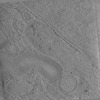 | |||||||||
|---|---|---|---|---|---|---|---|---|---|---|
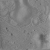
| Title | In situ cryo-electron tomogram of a multi-lamellar vesicle in a NPC2-/- HeLa cell. #1 | |||||||||
 Map data Map data | Highly organized membrane structures with NPC2-/- MLVs | |||||||||
 Sample Sample |
| |||||||||
 Keywords Keywords | Lysosomal storage disorders / autophagy / Multi-lamellar vesicle / cholesterol export / endocytosis / cargo delivery / LIPID TRANSPORT | |||||||||
| Biological species |  Homo sapiens (human) Homo sapiens (human) | |||||||||
| Method | electron tomography / cryo EM | |||||||||
 Authors Authors | Kraus F / He Y / Swarup S / Overmyer KA / Jiang Y / Brenner J / Capitanio C / Bieber A / Jen A / Nightingale NM ...Kraus F / He Y / Swarup S / Overmyer KA / Jiang Y / Brenner J / Capitanio C / Bieber A / Jen A / Nightingale NM / Anderson BJ / Lee C / Paulo JA / Smith IR / Plitzko JM / Gygi SP / Schulman BA / Wilfling F / Coon JJ / Harper JW | |||||||||
| Funding support |  United States, 1 items United States, 1 items
| |||||||||
 Citation Citation | Journal: Sci Adv / Year: 2025 Title: Global cellular proteo-lipidomic profiling of diverse lysosomal storage disease mutants using nMOST. Authors: Felix Kraus / Yuchen He / Sharan Swarup / Katherine A Overmyer / Yizhi Jiang / Johann Brenner / Cristina Capitanio / Anna Bieber / Annie Jen / Nicole M Nightingale / Benton J Anderson / Chan ...Authors: Felix Kraus / Yuchen He / Sharan Swarup / Katherine A Overmyer / Yizhi Jiang / Johann Brenner / Cristina Capitanio / Anna Bieber / Annie Jen / Nicole M Nightingale / Benton J Anderson / Chan Lee / Joao A Paulo / Ian R Smith / Jürgen M Plitzko / Steven P Gygi / Brenda A Schulman / Florian Wilfling / Joshua J Coon / J Wade Harper /  Abstract: Lysosomal storage diseases (LSDs) comprise ~50 monogenic disorders marked by the buildup of cellular material in lysosomes, yet systematic global molecular phenotyping of proteins and lipids is ...Lysosomal storage diseases (LSDs) comprise ~50 monogenic disorders marked by the buildup of cellular material in lysosomes, yet systematic global molecular phenotyping of proteins and lipids is lacking. We present a nanoflow-based multiomic single-shot technology (nMOST) workflow that quantifies HeLa cell proteomes and lipidomes from over two dozen LSD mutants. Global cross-correlation analysis between lipids and proteins identified autophagy defects, notably the accumulation of ferritinophagy substrates and receptors, especially in and mutants, where lysosomes accumulate cholesterol. Autophagic and endocytic cargo delivery failures correlated with elevated lysophosphatidylcholine species and multilamellar structures visualized by cryo-electron tomography. Loss of mitochondrial cristae, MICOS complex components, and OXPHOS components rich in iron-sulfur cluster proteins in cells was largely alleviated when iron was provided through the transferrin system. This study reveals how lysosomal dysfunction affects mitochondrial homeostasis and underscores nMOST as a valuable discovery tool for identifying molecular phenotypes across LSDs. | |||||||||
| History |
|
- Structure visualization
| Supplemental images |
|---|

- Downloads & links
-EMDB archive
| Map data | emd_51701.map.gz | 523 MB |  EMDB map data format EMDB map data format | |
|---|---|---|---|---|
| Header (meta data) | emd-51701-v30.xmlemd-51701.xml | 12.8 KB 12.8 KB | Display Display | EMDB header |
| Images |  emd_51701.png emd_51701.png | 213 KB | ||
| Filedesc metadata | emd-51701.cif.gz | 4.8 KB | ||
| Archive directory |  http://ftp.pdbj.org/pub/emdb/structures/EMD-51701ftp://ftp.pdbj.org/pub/emdb/structures/EMD-51701 http://ftp.pdbj.org/pub/emdb/structures/EMD-51701ftp://ftp.pdbj.org/pub/emdb/structures/EMD-51701 | HTTPS FTP |
-Related structure data
-Links
| EMDB pages | EMDB (EBI/PDBe) / EMDataResource |
|---|
-Map
| File | Download / File: emd_51701.map.gz / Format: CCP4 / Size: 564 MB / Type: IMAGE STORED AS FLOATING POINT NUMBER (4 BYTES) | ||||||||||||||||||||||||||||||||
|---|---|---|---|---|---|---|---|---|---|---|---|---|---|---|---|---|---|---|---|---|---|---|---|---|---|---|---|---|---|---|---|---|---|
| Annotation | Highly organized membrane structures with NPC2-/- MLVs | ||||||||||||||||||||||||||||||||
| Projections & slices | Image control
Images are generated by Spider. generated in cubic-lattice coordinate | ||||||||||||||||||||||||||||||||
| Voxel size | X=Y=Z: 11.72 Å | ||||||||||||||||||||||||||||||||
| Density |
| ||||||||||||||||||||||||||||||||
| Symmetry | Space group: 1 | ||||||||||||||||||||||||||||||||
| Details | EMDB XML:
|
 Z (Sec.)
 Y (Row.)
 X (Col.)


-Supplemental data
- Sample components
-Entire : HeLa TMEM192-3xHA NPC2-/-
| Entire | Name: HeLa TMEM192-3xHA NPC2-/- |
|---|---|
| Components |
|
-Supramolecule #1: HeLa TMEM192-3xHA NPC2-/-
| Supramolecule | Name: HeLa TMEM192-3xHA NPC2-/- / type: cell / ID: 1 / Parent: 0 Details: Modified with CRISPR/CAS9 with target sites determined using CHOPCHOP. |
|---|---|
| Source (natural) | Organism: Homo sapiens (human) |
-Experimental details
-Structure determination
| Method | cryo EM |
|---|---|
 Processing Processing | electron tomography |
| Aggregation state | cell |
-Sample preparation
| Buffer | pH: 7 |
|---|---|
| Grid | Model: Quantifoil R1/4 / Material: GOLD / Mesh: 200 / Support film - topology: HOLEY / Support film - Film thickness: 10 / Pretreatment - Type: GLOW DISCHARGE / Pretreatment - Time: 60 sec. / Pretreatment - Atmosphere: AIR |
| Vitrification | Cryogen name: ETHANE-PROPANE / Chamber humidity: 70 % / Chamber temperature: 298 K / Instrument: FEI VITROBOT MARK IV |
| Details | HeLa (TMEM192-HA) NPC2-/- cells were cultured on Poly-L-Lysine coated gold grids over night. The next day, cells were starved for 6h in EBSS. 10% glycerol was added shortly before plunging. |
| Cryo protectant | 10% glycerol |
| Sectioning | Focused ion beam - Instrument: OTHER / Focused ion beam - Ion: OTHER / Focused ion beam - Voltage: 30 / Focused ion beam - Current: 3 / Focused ion beam - Duration: 120 / Focused ion beam - Temperature: 80 K / Focused ion beam - Initial thickness: 1000 / Focused ion beam - Final thickness: 130 Focused ion beam - Details: The value given for _em_focused_ion_beam.instrument is Thermo Fisher Arctis PFIB. This is not in a list of allowed values {'DB235', 'OTHER'} so OTHER is written into the XML file. |
- Electron microscopy
| Microscope | TFS KRIOS |
|---|---|
| Specialist optics | Energy filter - Name: TFS Selectris X / Energy filter - Slit width: 10 eV |
| Image recording | Film or detector model: FEI FALCON IV (4k x 4k) / Digitization - Dimensions - Width: 4096 pixel / Digitization - Dimensions - Height: 4096 pixel / Average electron dose: 2.0 e/Å2 |
| Electron beam | Acceleration voltage: 300 kV / Electron source:  FIELD EMISSION GUN FIELD EMISSION GUN |
| Electron optics | Illumination mode: FLOOD BEAM / Imaging mode: BRIGHT FIELD / Nominal defocus max: 5.5 µm / Nominal defocus min: 3.5 µm / Nominal magnification: 42000 |
| Sample stage | Specimen holder model: FEI TITAN KRIOS AUTOGRID HOLDER / Cooling holder cryogen: NITROGEN |
| Experimental equipment |  Model: Titan Krios / Image courtesy: FEI Company |
-Image processing
| Final reconstruction | Algorithm: BACK PROJECTION / Software - Version: 1.3 / Software - details: Aretomo / Number images used: 47 |
|---|
