 Movie
Movie Controller


+ Open data

- Basic information
| Entry |  | |||||||||||||||||||||
|---|---|---|---|---|---|---|---|---|---|---|---|---|---|---|---|---|---|---|---|---|---|---|
| Title | Giant Jyvaskylavirus at 6.3 Ang resolution | |||||||||||||||||||||
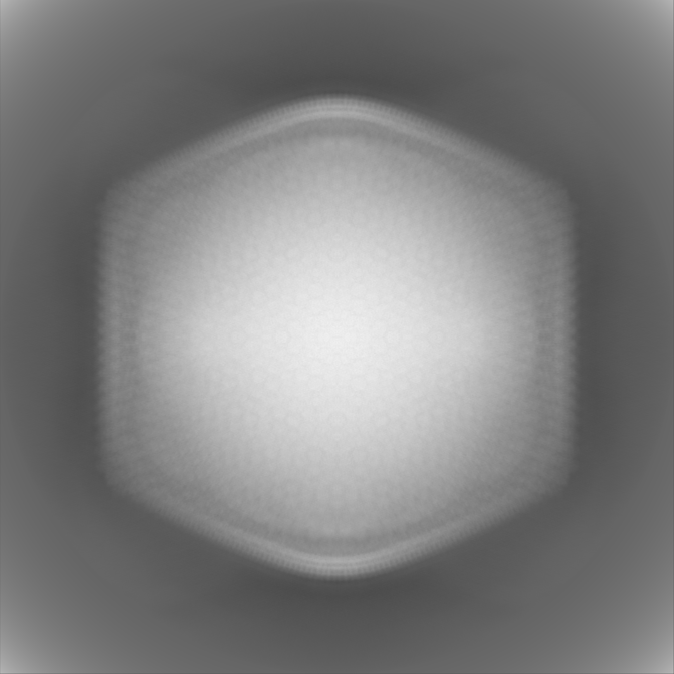
 Map data Map data | IMPORTANT: Map has been binned from the original boxel size (1.34 A/px). Post-processed map, no masked, using the automatic B-factor sharpening by RELION. | |||||||||||||||||||||
 Sample Sample |
| |||||||||||||||||||||
 Keywords Keywords | Giant virus / cryo-EM / genomic / NCLDV / Marseilleviridae family / VIRUS | |||||||||||||||||||||
| Biological species |  Acanthamoeba castellanii (eukaryote) / Acanthamoeba castellanii (eukaryote) /  Jyvaskylavirus Jyvaskylavirus | |||||||||||||||||||||
| Method | single particle reconstruction / cryo EM / Resolution: 6.3 Å | |||||||||||||||||||||
 Authors Authors | Abrescia NG / Arriaga I / de Freitas Almeida GM / Sundberg LR / Ravantti J | |||||||||||||||||||||
| Funding support |  Spain, Spain,  Finland, Finland,  Norway, Norway,  United Kingdom, 6 items United Kingdom, 6 items
| |||||||||||||||||||||
 Citation Citation | Journal: Elife / Year: 2025 Title: Genomic and structural insights into Jyvaskylavirus, the first giant virus isolated from Finland. Authors: Gabriel Magno de Freitas Almeida / Iker Arriaga / Bruna Luiza de Azevedo / Miika Leppänen / Jonatas S Abrahão / Julien Andreani / Davide Zabeo / Janne J Ravantti / Nicola G A Abrescia / ...Authors: Gabriel Magno de Freitas Almeida / Iker Arriaga / Bruna Luiza de Azevedo / Miika Leppänen / Jonatas S Abrahão / Julien Andreani / Davide Zabeo / Janne J Ravantti / Nicola G A Abrescia / Lotta-Riina Sundberg /   Abstract: Giant viruses of protists are a diverse and likely ubiquitous group of organisms. Here, we describe Jyvaskylavirus, the first giant virus isolated from Finland. This clade B marseillevirus was found ...Giant viruses of protists are a diverse and likely ubiquitous group of organisms. Here, we describe Jyvaskylavirus, the first giant virus isolated from Finland. This clade B marseillevirus was found in from a composting soil sample in Jyväskylä, Central Finland. Its genome shares similarities with other marseilleviruses. Helium ion microscopy and electron microscopy of infected cells unraveled stages of the Jyvaskylavirus life cycle. We reconstructed the Jyvaskylavirus particle to 6.3 Å resolution using cryo-electron microscopy. The ~2500 Å diameter virion displays structural similarities to other Marseilleviridae giant viruses. The capsid comprises of 9240 copies of the major capsid protein, encoded by open reading frame (ORF) 184, which possesses a double jellyroll fold arranged in trimers forming pseudo-hexameric capsomers. Below the capsid shell, the internal membrane vesicle encloses the genome. Through cross-structural and -sequence comparisons with other Marseilleviridae using AI-based software in model building and prediction, we elucidated ORF142 as the penton protein, which plugs the 12 vertices of the capsid. Five additional ORFs were identified, with models predicted and fitted into densities that either cap the capsomers externally or stabilize them internally. The isolation of Jyvaskylavirus suggests that these viruses may be widespread in the boreal environment and provide structural insights extendable to other marseilleviruses. | |||||||||||||||||||||
| History |
|
- Structure visualization
| Supplemental images |
|---|

- Downloads & links
-EMDB archive
| Map data | emd_51613.map.gz | 41.1 GB |  EMDB map data format EMDB map data format | |
|---|---|---|---|---|
| Header (meta data) | emd-51613-v30.xmlemd-51613.xml | 20.8 KB 20.8 KB | Display Display | EMDB header |
| FSC (resolution estimation) | emd_51613_fsc.xml | 81.5 KB | Display | FSC data file |
| Images |  emd_51613.png emd_51613.png | 229.9 KB | ||
| Filedesc metadata | emd-51613.cif.gz | 5.8 KB | ||
| Others | emd_51613_half_map_1.map.gzemd_51613_half_map_2.map.gz | 37.6 GB 37.6 GB | ||
| Archive directory |  http://ftp.pdbj.org/pub/emdb/structures/EMD-51613ftp://ftp.pdbj.org/pub/emdb/structures/EMD-51613 http://ftp.pdbj.org/pub/emdb/structures/EMD-51613ftp://ftp.pdbj.org/pub/emdb/structures/EMD-51613 | HTTPS FTP |
-Links
| EMDB pages | EMDB (EBI/PDBe) / EMDataResource |
|---|
-Map
| File | Download / File: emd_51613.map.gz / Format: CCP4 / Size: 5.7 GB / Type: IMAGE STORED AS FLOATING POINT NUMBER (4 BYTES) | ||||||||||||||||||||||||||||||||
|---|---|---|---|---|---|---|---|---|---|---|---|---|---|---|---|---|---|---|---|---|---|---|---|---|---|---|---|---|---|---|---|---|---|
| Annotation | IMPORTANT: Map has been binned from the original boxel size (1.34 A/px). Post-processed map, no masked, using the automatic B-factor sharpening by RELION. | ||||||||||||||||||||||||||||||||
| Projections & slices | Image control
Images are generated by Spider. | ||||||||||||||||||||||||||||||||
| Voxel size | X=Y=Z: 1.34 Å | ||||||||||||||||||||||||||||||||
| Density |
| ||||||||||||||||||||||||||||||||
| Symmetry | Space group: 1 | ||||||||||||||||||||||||||||||||
| Details | EMDB XML:
|
 Z (Sec.)
 Y (Row.)
 X (Col.)


-Supplemental data
-Half map: IMPORTANT: Map has been binned from the original...
| File | emd_51613_half_map_1.map | ||||||||||||
|---|---|---|---|---|---|---|---|---|---|---|---|---|---|
| Annotation | IMPORTANT: Map has been binned from the original boxel size (1.34 A/px). Unfiltered, unmasked, half map 2 from the 3D auto refinement by RELION | ||||||||||||
| Projections & Slices |
| ||||||||||||
| Density Histograms |


-Half map: IMPORTANT: Map has been binned from the original...
| File | emd_51613_half_map_2.map | ||||||||||||
|---|---|---|---|---|---|---|---|---|---|---|---|---|---|
| Annotation | IMPORTANT: Map has been binned from the original boxel size (1.34 A/px). Unfiltered, unmasked, half map 1 from the 3D auto refinement by RELION | ||||||||||||
| Projections & Slices |
| ||||||||||||
| Density Histograms |


- Sample components
-Entire : Jyvaskylavirus
| Entire | Name: Jyvaskylavirus |
|---|---|
| Components |
|
-Supramolecule #1: Jyvaskylavirus
| Supramolecule | Name: Jyvaskylavirus / type: virus / ID: 1 / Parent: 0 / Macromolecule list: all / Details: Full virion / NCBI-ID: 1513458 / Sci species name: Jyvaskylavirus / Virus type: VIRION / Virus isolate: OTHER / Virus enveloped: No / Virus empty: No |
|---|---|
| Host (natural) | Organism: Acanthamoeba castellanii (eukaryote) |
| Virus shell | Shell ID: 1 / Name: Jyvaskylavirus ORF184 / Diameter: 2500.0 Å / T number (triangulation number): 309 |
-Macromolecule #1: Jyvaskylavirus
| Macromolecule | Name: Jyvaskylavirus / type: other / ID: 1 / Classification: other |
|---|---|
| Source (natural) | Organism: Acanthamoeba castellanii (eukaryote) |
| Sequence | String: XXXXX |
-Experimental details
-Structure determination
| Method | cryo EM |
|---|---|
 Processing Processing | single particle reconstruction |
| Aggregation state | particle |
-Sample preparation
| Buffer | pH: 7.4 Component:
| ||||||||||||||||||
|---|---|---|---|---|---|---|---|---|---|---|---|---|---|---|---|---|---|---|---|
| Grid | Model: Quantifoil R2/1 / Material: COPPER / Mesh: 300 / Support film - #0 - Film type ID: 1 / Support film - #0 - Material: CARBON / Support film - #0 - topology: HOLEY / Support film - #1 - Film type ID: 2 / Support film - #1 - Material: CARBON / Support film - #1 - topology: CONTINUOUS / Pretreatment - Type: GLOW DISCHARGE / Pretreatment - Time: 37 sec. / Pretreatment - Atmosphere: AIR / Pretreatment - Pressure: 0.0002 kPa / Details: 8-9 mA | ||||||||||||||||||
| Vitrification | Cryogen name: ETHANE / Chamber humidity: 95 % / Chamber temperature: 277.15 K / Instrument: FEI VITROBOT MARK IV Details: Also Automatic Plunge Freezer EM2 (Leica) was used (see publication).. |
- Electron microscopy
| Microscope | TFS KRIOS |
|---|---|
| Image recording | Film or detector model: GATAN K3 BIOQUANTUM (6k x 4k) / Average electron dose: 45.0 e/Å2 |
| Electron beam | Acceleration voltage: 300 kV / Electron source:  FIELD EMISSION GUN FIELD EMISSION GUN |
| Electron optics | Illumination mode: FLOOD BEAM / Imaging mode: BRIGHT FIELD / Nominal defocus max: 3.0 µm / Nominal defocus min: 0.6 µm / Nominal magnification: 64000 |
| Experimental equipment |  Model: Titan Krios / Image courtesy: FEI Company |
+Image processing

-Atomic model buiding 1
| Initial model |
| ||||||||
|---|---|---|---|---|---|---|---|---|---|
| Details | Please keep in mind that the predicted Jyvaskylavirus PDB models using ModelAngelo and Alphafold have been deposited at the Biostudies website (https://www.ebi.ac.uk/biostudies/). Please refer to the published article for the corresponding entry code. | ||||||||
| Refinement | Protocol: RIGID BODY FIT |
