Biotechnology and Biological Sciences Research Council (BBSRC)
BB/R009759/2
United Kingdom
Citation
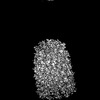
Journal: Nat Microbiol / Year: 2025 Title: The structure of the complete extracellular bacterial flagellum reveals the mechanism of flagellin incorporation. Authors: Rosa Einenkel / Kailin Qin / Julia Schmidt / Natalie S Al-Otaibi / Daniel Mann / Tina Drobnič / Eli J Cohen / Nayim Gonzalez-Rodriguez / Jane Harrowell / Elena Shmakova / Morgan Beeby / ...Authors: Rosa Einenkel / Kailin Qin / Julia Schmidt / Natalie S Al-Otaibi / Daniel Mann / Tina Drobnič / Eli J Cohen / Nayim Gonzalez-Rodriguez / Jane Harrowell / Elena Shmakova / Morgan Beeby / Marc Erhardt / Julien R C Bergeron / Abstract: The bacterial flagellum is essential for motility, adhesion and colonization in pathogens such as Salmonella enterica and Campylobacter jejuni. Its extracellular structure comprises the hook, hook- ...The bacterial flagellum is essential for motility, adhesion and colonization in pathogens such as Salmonella enterica and Campylobacter jejuni. Its extracellular structure comprises the hook, hook-filament junction, filament and filament cap. Native structures of the hook-filament junction and the cap are lacking, and molecular mechanisms of cap-mediated filament assembly are largely uncharacterized. Here we use cryo-electron microscopy to resolve structures of the complete Salmonella extracellular flagellum including the pentameric FliD cap complex (3.7 Å) and the FlgKL hook-filament junction (2.9 Å), as well as the Campylobacter extracellular flagellum before filament assembly (6.5 Å). This, coupled with structure-guided mutagenesis and functional assays, reveals intermediates of filament assembly, showing that FliD cap protein terminal domain movement and clockwise rotation enable flagellin incorporation and stabilization of the filament. We show that the hook-filament junction acts as a buffer, preventing transfer of mechanical stress to the filament, and reveal the structural basis for the initiation of filament assembly. Collectively, this study provides comprehensive insights into flagellum assembly and how flagellin incorporation is coupled with its secretion.
In the structure databanks used in Yorodumi, some data are registered as the other names, "COVID-19 virus" and "2019-nCoV". Here are the details of the virus and the list of structure data.
Jan 31, 2019. EMDB accession codes are about to change! (news from PDBe EMDB page)
EMDB accession codes are about to change! (news from PDBe EMDB page)
The allocation of 4 digits for EMDB accession codes will soon come to an end. Whilst these codes will remain in use, new EMDB accession codes will include an additional digit and will expand incrementally as the available range of codes is exhausted. The current 4-digit format prefixed with “EMD-” (i.e. EMD-XXXX) will advance to a 5-digit format (i.e. EMD-XXXXX), and so on. It is currently estimated that the 4-digit codes will be depleted around Spring 2019, at which point the 5-digit format will come into force.
The EM Navigator/Yorodumi systems omit the EMD- prefix.
Related info.:Q: What is EMD? / ID/Accession-code notation in Yorodumi/EM Navigator
Yorodumi is a browser for structure data from EMDB, PDB, SASBDB, etc.
This page is also the successor to EM Navigator detail page, and also detail information page/front-end page for Omokage search.
The word "yorodu" (or yorozu) is an old Japanese word meaning "ten thousand". "mi" (miru) is to see.
Related info.:EMDB / PDB / SASBDB / Comparison of 3 databanks / Yorodumi Search / Aug 31, 2016. New EM Navigator & Yorodumi / Yorodumi Papers / Jmol/JSmol / Function and homology information / Changes in new EM Navigator and Yorodumi
 Movie
Movie Controller


 Open data
 Basic information
 Map data
 Sample
 Keywords
 Salmonella enterica (bacteria)
 Authors
 United Kingdom, 1 items
  Citation

 Structure visualization
 Downloads & links
 EMDB map data format
 emd_51555.png
 http://ftp.pdbj.org/pub/emdb/structures/EMD-51555


 Z (Sec.)
 Y (Row.)
 X (Col.)


 Sample components
 Processing
 Electron microscopy
 FIELD EMISSION GUN
