 Movie
Movie Controller

[English] 日本語
 Yorodumi
- EMDB-51521: Cryo-electron microscopy structure of glucose/xylose isomerase fr... -
+ Open data

- Basic information
| Entry |  | |||||||||
|---|---|---|---|---|---|---|---|---|---|---|
| Title | Cryo-electron microscopy structure of glucose/xylose isomerase from Streptomyces rubiginosus with cobalt ions in the active site | |||||||||
 Map data Map data | Volume map | |||||||||
 Sample Sample |
| |||||||||
 Keywords Keywords | cobalt / isomerase / sugars / glucose / xylose / metal ion / Cryo-EM / METAL BINDING PROTEIN | |||||||||
| Function / homology |  Function and homology information Function and homology informationxylose isomerase / xylose isomerase activity / D-xylose metabolic process / magnesium ion binding / identical protein binding / cytoplasm Similarity search - Function | |||||||||
| Biological species |  Streptomyces rubiginosus (bacteria) Streptomyces rubiginosus (bacteria) | |||||||||
| Method | single particle reconstruction / cryo EM / Resolution: 1.99 Å | |||||||||
 Authors Authors | Slawek J / Klonecka A / Rawski M / Kozak M | |||||||||
| Funding support |  Poland, 1 items Poland, 1 items
| |||||||||
 Citation Citation | Journal: To Be Published Title: Cryo-electron microscopy structure of glucose/xylose isomerase from Streptomyces rubiginosus with cobalt ions in the active site Authors: Slawek J / Klonecka A / Kozak M / Rawski M | |||||||||
| History |
|
- Structure visualization
| Supplemental images |
|---|

- Downloads & links
-EMDB archive
| Map data | emd_51521.map.gz | 31.5 MB | EMDB map data format | |
|---|---|---|---|---|
| Header (meta data) | emd-51521-v30.xmlemd-51521.xml | 18.2 KB 18.2 KB | Display Display | EMDB header |
| FSC (resolution estimation) | emd_51521_fsc.xml | 11.6 KB | Display | FSC data file |
| Images |  emd_51521.png emd_51521.png | 103.7 KB | ||
| Filedesc metadata | emd-51521.cif.gz | 5.7 KB | ||
| Others | emd_51521_additional_1.map.gzemd_51521_half_map_1.map.gzemd_51521_half_map_2.map.gz | 56.9 MB 59.1 MB 59.1 MB | ||
| Archive directory |  http://ftp.pdbj.org/pub/emdb/structures/EMD-51521ftp://ftp.pdbj.org/pub/emdb/structures/EMD-51521 http://ftp.pdbj.org/pub/emdb/structures/EMD-51521ftp://ftp.pdbj.org/pub/emdb/structures/EMD-51521 | HTTPS FTP |
-Related structure data
| Related structure data |  9grdMC M: atomic model generated by this map C: citing same article ( |
|---|---|
| Similar structure data |
-Links
| EMDB pages | EMDB (EBI/PDBe) / EMDataResource |
|---|---|
| Related items in Molecule of the Month |

-Map
| File | Download / File: emd_51521.map.gz / Format: CCP4 / Size: 64 MB / Type: IMAGE STORED AS FLOATING POINT NUMBER (4 BYTES) | ||||||||||||||||||||||||||||||||||||
|---|---|---|---|---|---|---|---|---|---|---|---|---|---|---|---|---|---|---|---|---|---|---|---|---|---|---|---|---|---|---|---|---|---|---|---|---|---|
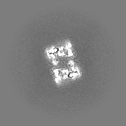
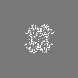
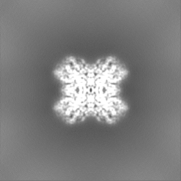
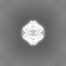
| Annotation | Volume map | ||||||||||||||||||||||||||||||||||||
| Projections & slices | Image control
Images are generated by Spider. | ||||||||||||||||||||||||||||||||||||
| Voxel size | X=Y=Z: 0.84 Å | ||||||||||||||||||||||||||||||||||||
| Density |
| ||||||||||||||||||||||||||||||||||||
| Symmetry | Space group: 1 | ||||||||||||||||||||||||||||||||||||
| Details | EMDB XML:
|
 Z (Sec.)
 Y (Row.)
 X (Col.)


-Supplemental data
-Additional map: map sharp
| File | emd_51521_additional_1.map | ||||||||||||
|---|---|---|---|---|---|---|---|---|---|---|---|---|---|
| Annotation | map sharp | ||||||||||||
| Projections & Slices |
| ||||||||||||
| Density Histograms |


-Half map: Half map B
| File | emd_51521_half_map_1.map | ||||||||||||
|---|---|---|---|---|---|---|---|---|---|---|---|---|---|
| Annotation | Half map B | ||||||||||||
| Projections & Slices |
| ||||||||||||
| Density Histograms |


-Half map: Half map A
| File | emd_51521_half_map_2.map | ||||||||||||
|---|---|---|---|---|---|---|---|---|---|---|---|---|---|
| Annotation | Half map A | ||||||||||||
| Projections & Slices |
| ||||||||||||
| Density Histograms |


- Sample components
-Entire : glucose/xylose isomerase with cobalt ions
| Entire | Name: glucose/xylose isomerase with cobalt ions |
|---|---|
| Components |
|
-Supramolecule #1: glucose/xylose isomerase with cobalt ions
| Supramolecule | Name: glucose/xylose isomerase with cobalt ions / type: complex / ID: 1 / Parent: 0 / Macromolecule list: #1 |
|---|---|
| Source (natural) | Organism: Streptomyces rubiginosus (bacteria) |
| Molecular weight | Theoretical: 43 KDa |
-Macromolecule #1: Xylose isomerase
| Macromolecule | Name: Xylose isomerase / type: protein_or_peptide / ID: 1 / Number of copies: 4 / Enantiomer: LEVO / EC number: xylose isomerase |
|---|---|
| Source (natural) | Organism: Streptomyces rubiginosus (bacteria) |
| Molecular weight | Theoretical: 43.283297 KDa |
| Sequence | String: MNYQPTPEDR FTFGLWTVGW QGRDPFGDAT RRALDPVESV RRLAELGAHG VTFHDDDLIP FGSSDSEREE HVKRFRQALD DTGMKVPMA TTNLFTHPVF KDGGFTANDR DVRRYALRKT IRNIDLAVEL GAETYVAWGG REGAESGGAK DVRDALDRMK E AFDLLGEY ...String: MNYQPTPEDR FTFGLWTVGW QGRDPFGDAT RRALDPVESV RRLAELGAHG VTFHDDDLIP FGSSDSEREE HVKRFRQALD DTGMKVPMA TTNLFTHPVF KDGGFTANDR DVRRYALRKT IRNIDLAVEL GAETYVAWGG REGAESGGAK DVRDALDRMK E AFDLLGEY VTSQGYDIRF AIEPKPNEPR GDILLPTVGH ALAFIERLER PELYGVNPEV GHEQMAGLNF PHGIAQALWA GK LFHIDLN GQNGIKYDQD LRFGAGDLRA AFWLVDLLES AGYSGPRHFD FKPPRTEDFD GVWASAAGCM RNYLILKERA AAF RADPEV QEALRASRLD ELARPTAADG LQALLDDRSA FEEFDVDAAA ARGMAFERLD QLAMDHLLGA RG UniProtKB: Xylose isomerase |
-Macromolecule #2: COBALT (II) ION
| Macromolecule | Name: COBALT (II) ION / type: ligand / ID: 2 / Number of copies: 8 / Formula: CO |
|---|---|
| Molecular weight | Theoretical: 58.933 Da |
-Macromolecule #3: water
| Macromolecule | Name: water / type: ligand / ID: 3 / Number of copies: 4 / Formula: HOH |
|---|---|
| Molecular weight | Theoretical: 18.015 Da |
| Chemical component information |  ChemComp-HOH: |
-Experimental details
-Structure determination
| Method | cryo EM |
|---|---|
 Processing Processing | single particle reconstruction |
| Aggregation state | 3D array |
-Sample preparation
| Concentration | 1.04 mg/mL |
|---|---|
| Buffer | pH: 7.4 / Component - Concentration: 50.0 mM / Component - Formula: PBS / Component - Name: Phosphate-buffered saline / Details: 50 mM PBS pH 7.4 |
| Grid | Model: Quantifoil R2/1 / Material: COPPER / Mesh: 200 / Pretreatment - Type: GLOW DISCHARGE / Pretreatment - Time: 5 sec. / Pretreatment - Atmosphere: AIR |
| Vitrification | Cryogen name: ETHANE |
- Electron microscopy
| Microscope | TFS KRIOS |
|---|---|
| Specialist optics | Energy filter - Name: GIF Bioquantum / Energy filter - Slit width: 20 eV |
| Image recording | Film or detector model: GATAN K3 BIOQUANTUM (6k x 4k) / Average electron dose: 40.0 e/Å2 |
| Electron beam | Acceleration voltage: 300 kV / Electron source:  FIELD EMISSION GUN FIELD EMISSION GUN |
| Electron optics | Illumination mode: FLOOD BEAM / Imaging mode: BRIGHT FIELD / Nominal defocus max: 2.4 µm / Nominal defocus min: 0.9 µm / Nominal magnification: 105000 |
| Sample stage | Specimen holder model: FEI TITAN KRIOS AUTOGRID HOLDER / Cooling holder cryogen: NITROGEN |
| Experimental equipment |  Model: Titan Krios / Image courtesy: FEI Company |

