 Movie
Movie Controller

[English] 日本語
 Yorodumi
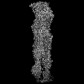
- EMDB-51491: CryoEM Reconstruction of Yeast ADP-Actin Filament at 2.5 A resolution. -
+ Open data

- Basic information
| Entry |  | |||||||||
|---|---|---|---|---|---|---|---|---|---|---|
| Title | CryoEM Reconstruction of Yeast ADP-Actin Filament at 2.5 A resolution. | |||||||||
 Map data Map data | Sharpened CryoEM map of yeast actin | |||||||||
 Sample Sample |
| |||||||||
 Keywords Keywords | Fibre / polymer / helical protein / motility / PROTEIN FIBRIL | |||||||||
| Function / homology |  Function and homology information Function and homology informationcellular bud neck contractile ring / mitotic actomyosin contractile ring contraction / : / RHOA GTPase cycle / vacuole inheritance / ascospore wall assembly / actin cortical patch / Swr1 complex / Ino80 complex / establishment of cell polarity ...cellular bud neck contractile ring / mitotic actomyosin contractile ring contraction / : / RHOA GTPase cycle / vacuole inheritance / ascospore wall assembly / actin cortical patch / Swr1 complex / Ino80 complex / establishment of cell polarity / actin filament bundle / NuA4 histone acetyltransferase complex / protein secretion / actin filament / structural constituent of cytoskeleton / Hydrolases; Acting on acid anhydrides; Acting on acid anhydrides to facilitate cellular and subcellular movement / endocytosis / actin cytoskeleton / chromatin remodeling / DNA repair / regulation of DNA-templated transcription / chromatin / DNA-templated transcription / ATP hydrolysis activity / ATP binding / identical protein binding / nucleus Similarity search - Function | |||||||||
| Biological species |  | |||||||||
| Method | helical reconstruction / cryo EM / Resolution: 2.5 Å | |||||||||
 Authors Authors | Bullough PA / Ayscough KR / Lahiri I / Tzokov SB / Stevenson SR | |||||||||
| Funding support |  United Kingdom, 1 items United Kingdom, 1 items
| |||||||||
 Citation Citation | Journal: Structure / Year: 2025 Title: Cryo-EM reconstruction of yeast ADP-actin filament at 2.5 Å resolution. A comparison with vertebrate F-actin. Authors: Sarah R Stevenson / Svetomir B Tzokov / Indrajit Lahiri / Kathryn R Ayscough / Per A Bullough / Abstract: The core component of the actin cytoskeleton is the globular protein G-actin, which reversibly polymerizes into filaments (F-actin). Budding yeast possesses a single actin that shares 87%-89% ...The core component of the actin cytoskeleton is the globular protein G-actin, which reversibly polymerizes into filaments (F-actin). Budding yeast possesses a single actin that shares 87%-89% sequence identity with vertebrate actin isoforms. Previous structural studies indicate very close overlap of main-chain backbones. Intriguingly, however, substitution of yeast ACT1 with vertebrate β-cytoplasmic actin severely disrupts cell function and the substitution with a skeletal muscle isoform is lethal. Here we report a 2.5 Å structure of budding yeast F-actin. Previously unresolved side-chain information allows us to highlight four main differences in the comparison of yeast and vertebrate ADP F-actins: a more open nucleotide binding pocket; a more solvent exposed C-terminus; a rearrangement of inter-subunit binding interactions in the vicinity of the D loop and changes in the hydrogen bonding network in the vicinity of histidine 73 (yeast actin) and methyl-histidine 73 (vertebrate actin). | |||||||||
| History |
|
- Structure visualization
| Supplemental images |
|---|

- Downloads & links
-EMDB archive
| Map data | emd_51491.map.gz | 47.3 MB | EMDB map data format | |
|---|---|---|---|---|
| Header (meta data) | emd-51491-v30.xmlemd-51491.xml | 17.1 KB 17.1 KB | Display Display | EMDB header |
| FSC (resolution estimation) | emd_51491_fsc.xml | 9.5 KB | Display | FSC data file |
| Images |  emd_51491.png emd_51491.png | 35.1 KB | ||
| Filedesc metadata | emd-51491.cif.gz | 6 KB | ||
| Others | emd_51491_half_map_1.map.gzemd_51491_half_map_2.map.gz | 84.4 MB 84.4 MB | ||
| Archive directory |  http://ftp.pdbj.org/pub/emdb/structures/EMD-51491ftp://ftp.pdbj.org/pub/emdb/structures/EMD-51491 http://ftp.pdbj.org/pub/emdb/structures/EMD-51491ftp://ftp.pdbj.org/pub/emdb/structures/EMD-51491 | HTTPS FTP |
-Related structure data
| Related structure data |  9go5MC M: atomic model generated by this map C: citing same article ( |
|---|---|
| Similar structure data |
-Links
| EMDB pages | EMDB (EBI/PDBe) / EMDataResource |
|---|---|
| Related items in Molecule of the Month |


-Map
| File | Download / File: emd_51491.map.gz / Format: CCP4 / Size: 91.1 MB / Type: IMAGE STORED AS FLOATING POINT NUMBER (4 BYTES) | ||||||||||||||||||||||||||||||||||||
|---|---|---|---|---|---|---|---|---|---|---|---|---|---|---|---|---|---|---|---|---|---|---|---|---|---|---|---|---|---|---|---|---|---|---|---|---|---|
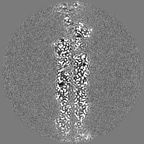
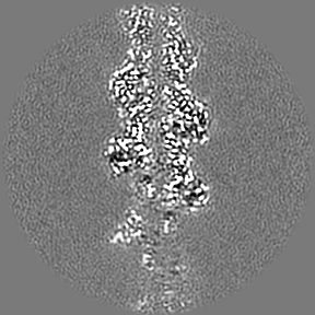
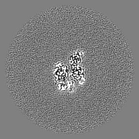
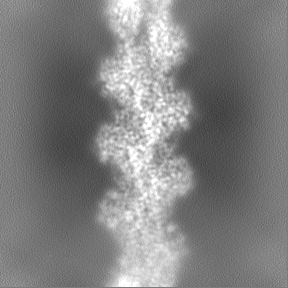
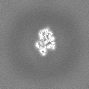
| Annotation | Sharpened CryoEM map of yeast actin | ||||||||||||||||||||||||||||||||||||
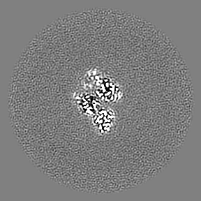
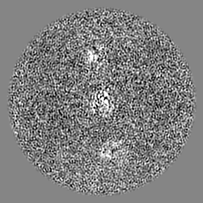
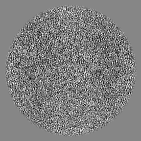
| Projections & slices | Image control
Images are generated by Spider. | ||||||||||||||||||||||||||||||||||||
| Voxel size | X=Y=Z: 0.834 Å | ||||||||||||||||||||||||||||||||||||
| Density |
| ||||||||||||||||||||||||||||||||||||
| Symmetry | Space group: 1 | ||||||||||||||||||||||||||||||||||||
| Details | EMDB XML:
|
 Z (Sec.)
 Y (Row.)
 X (Col.)


-Supplemental data
-Half map: CryoEM half map of yeast actin
| File | emd_51491_half_map_1.map | ||||||||||||
|---|---|---|---|---|---|---|---|---|---|---|---|---|---|
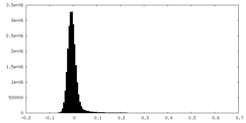
| Annotation | CryoEM half map of yeast actin | ||||||||||||
| Projections & Slices |
| ||||||||||||
| Density Histograms |


-Half map: CryoEM half map of yeast actin
| File | emd_51491_half_map_2.map | ||||||||||||
|---|---|---|---|---|---|---|---|---|---|---|---|---|---|
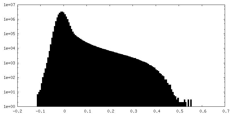
| Annotation | CryoEM half map of yeast actin | ||||||||||||
| Projections & Slices |
| ||||||||||||
| Density Histograms |


- Sample components
-Entire : Polymer of actin subunits
| Entire | Name: Polymer of actin subunits |
|---|---|
| Components |
|
-Supramolecule #1: Polymer of actin subunits
| Supramolecule | Name: Polymer of actin subunits / type: complex / ID: 1 / Parent: 0 / Macromolecule list: #1 |
|---|---|
| Source (natural) | Organism: |
-Macromolecule #1: Actin
| Macromolecule | Name: Actin / type: protein_or_peptide / ID: 1 / Number of copies: 5 / Enantiomer: LEVO EC number: Hydrolases; Acting on acid anhydrides; Acting on acid anhydrides to facilitate cellular and subcellular movement |
|---|---|
| Source (natural) | Organism: |
| Molecular weight | Theoretical: 41.735547 KDa |
| Recombinant expression | Organism:  Komagataella pastoris (fungus) Komagataella pastoris (fungus) |
| Sequence | String: MDSEVAALVI DNGSGMCKAG FAGDDAPRAV FPSIVGRPRH QGIMVGMGQK DSYVGDEAQS KRGILTLRYP IEHGIVTNWD DMEKIWHHT FYNELRVAPE EHPVLLTEAP MNPKSNREKM TQIMFETFNV PAFYVSIQAV LSLYSSGRTT GIVLDSGDGV T HVVPIYAG ...String: MDSEVAALVI DNGSGMCKAG FAGDDAPRAV FPSIVGRPRH QGIMVGMGQK DSYVGDEAQS KRGILTLRYP IEHGIVTNWD DMEKIWHHT FYNELRVAPE EHPVLLTEAP MNPKSNREKM TQIMFETFNV PAFYVSIQAV LSLYSSGRTT GIVLDSGDGV T HVVPIYAG FSLPHAILRI DLAGRDLTDY LMKILSERGY SFSTTAEREI VRDIKEKLCY VALDFEQEMQ TAAQSSSIEK SY ELPDGQV ITIGNERFRA PEALFHPSVL GLESAGIDQT TYNSIMKCDV DVRKELYGNI VMSGGTTMFP GIAERMQKEI TAL APSSMK VKIIAPPERK YSVWIGGSIL ASLTTFQQMW ISKQEYDESG PSIVHHKCF UniProtKB: Actin |
-Macromolecule #2: ADENOSINE-5'-DIPHOSPHATE
| Macromolecule | Name: ADENOSINE-5'-DIPHOSPHATE / type: ligand / ID: 2 / Number of copies: 5 / Formula: ADP |
|---|---|
| Molecular weight | Theoretical: 427.201 Da |
| Chemical component information |  ChemComp-ADP: |
-Macromolecule #3: MAGNESIUM ION
| Macromolecule | Name: MAGNESIUM ION / type: ligand / ID: 3 / Number of copies: 5 / Formula: MG |
|---|---|
| Molecular weight | Theoretical: 24.305 Da |
-Macromolecule #4: water
| Macromolecule | Name: water / type: ligand / ID: 4 / Number of copies: 350 / Formula: HOH |
|---|---|
| Molecular weight | Theoretical: 18.015 Da |
| Chemical component information |  ChemComp-HOH: |
-Experimental details
-Structure determination
| Method | cryo EM |
|---|---|
 Processing Processing | helical reconstruction |
| Aggregation state | helical array |
-Sample preparation
| Buffer | pH: 8 / Details: 50 mM KCl, 1 mM MgCl2, 1 mM EGTA, 10 mM Tris |
|---|---|
| Grid | Model: Quantifoil R2/2 / Material: COPPER / Mesh: 300 / Support film - Material: CARBON / Support film - topology: HOLEY / Pretreatment - Type: GLOW DISCHARGE |
| Vitrification | Cryogen name: ETHANE / Instrument: LEICA EM GP |
- Electron microscopy
| Microscope | FEI TITAN KRIOS |
|---|---|
| Image recording | Film or detector model: GATAN K3 (6k x 4k) / Detector mode: SUPER-RESOLUTION / Average electron dose: 27.0 e/Å2 |
| Electron beam | Acceleration voltage: 300 kV / Electron source:  FIELD EMISSION GUN FIELD EMISSION GUN |
| Electron optics | Illumination mode: FLOOD BEAM / Imaging mode: BRIGHT FIELD / Nominal defocus max: 2.4 µm / Nominal defocus min: 0.5 µm |
| Sample stage | Specimen holder model: FEI TITAN KRIOS AUTOGRID HOLDER / Cooling holder cryogen: NITROGEN |
| Experimental equipment |  Model: Titan Krios / Image courtesy: FEI Company |
-Image processing
| Final reconstruction | Applied symmetry - Helical parameters - Δz: 2.76 Å Applied symmetry - Helical parameters - Δ&Phi: -167.2 ° Applied symmetry - Helical parameters - Axial symmetry: C1 (asymmetric) Resolution.type: BY AUTHOR / Resolution: 2.5 Å / Resolution method: FSC 0.143 CUT-OFF / Number images used: 1368572 |
|---|---|
| Startup model | Type of model: NONE |
| Final angle assignment | Type: NOT APPLICABLE |
| FSC plot (resolution estimation) | 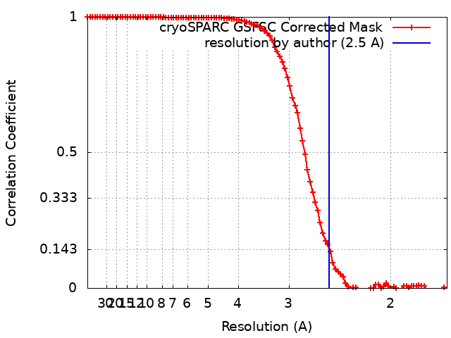 |
-Atomic model buiding 1
| Refinement | Space: REAL / Protocol: AB INITIO MODEL |
|---|---|
| Output model | PDB-9go5: |
