 Movie
Movie Controller

[English] 日本語
 Yorodumi
- EMDB-5147: Bluetongue virus structure reveals a sialic acid binding domain, ... -
+ Open data

- Basic information
| Entry | Database: EMDB / ID: EMD-5147 | |||||||||
|---|---|---|---|---|---|---|---|---|---|---|
| Title | Bluetongue virus structure reveals a sialic acid binding domain, amphipathic helices and a central coiled coil in the outer capsid proteins | |||||||||
 Map data Map data | BTV map | |||||||||
 Sample Sample |
| |||||||||
 Keywords Keywords | Bluetongue virus cryoEM sialic acid | |||||||||
| Function / homology |  Function and homology information Function and homology informationviral inner capsid / viral outer capsid / symbiont entry into host cell via permeabilization of host membrane / structural molecule activity Similarity search - Function | |||||||||
| Biological species |  Bluetongue virus Bluetongue virus | |||||||||
| Method | single particle reconstruction / cryo EM / negative staining / Resolution: 7.0 Å | |||||||||
 Authors Authors | Zhang X / Boyce M / Bhattacharya B / Schein S / Roy P / Zhou ZH | |||||||||
 Citation Citation | Journal: Proc Natl Acad Sci U S A / Year: 2010 Title: Bluetongue virus coat protein VP2 contains sialic acid-binding domains, and VP5 resembles enveloped virus fusion proteins. Authors: Xing Zhang / Mark Boyce / Bishnupriya Bhattacharya / Xiaokang Zhang / Stan Schein / Polly Roy / Z Hong Zhou /  Abstract: Bluetongue virus (BTV) is transmitted by blood-feeding insects (Culicoides sp.) and causes hemorrhagic diseases in livestock. BTV is a nonenveloped, double-stranded RNA (dsRNA) virus with two capsids: ...Bluetongue virus (BTV) is transmitted by blood-feeding insects (Culicoides sp.) and causes hemorrhagic diseases in livestock. BTV is a nonenveloped, double-stranded RNA (dsRNA) virus with two capsids: a well-studied, stable core enclosing the dsRNA genome and a highly unstable, poorly studied coat responsible for host cell attachment and entry. Here, based on cryo-electron microscopy (cryoEM), we report a 7-A resolution structure of the infectious BTV virion, including the coat proteins. We show that unlike other dsRNA viruses, the VP2 attachment trimer has a triskelion shape composed of three tip domains branching from a central hub domain. We identify three putative sialic acid-binding pockets in the hub and present supporting biochemical data indicating sugar moiety binding is important for BTV infection. Despite being a nonenveloped virus, the putative VP5 membrane penetration trimer, located slightly inward of the VP2 attachment trimer, has a central coiled-coil alpha-helical bundle, similar to the fusion proteins of many enveloped viruses (e.g., HIV, herpesviruses, vesicular stomatitis virus, and influenza virus). Moreover, mapping of the amino acid sequence of VP5 to the secondary structural elements identified by cryoEM locates 15 amphipathic alpha-helical regions on the external surface of each VP5 trimer. The cryoEM density map also reveals few, weak interactions between the VP5 trimer and both the outer-coat VP2 trimer and the underlying core VP7 trimer, suggesting that the surface of VP5 could unfurl like an umbrella during penetration and shedding of the coat to release the transcriptionally active core particle. | |||||||||
| History |
|
- Structure visualization
| Movie |
Movie viewer |
|---|---|
| Structure viewer | EM map: SurfViewMolmilJmol/JSmol |
| Supplemental images |

- Downloads & links
-EMDB archive
| Map data | emd_5147.map.gz | 403.5 MB | EMDB map data format | |
|---|---|---|---|---|
| Header (meta data) | emd-5147-v30.xmlemd-5147.xml | 13.5 KB 13.5 KB | Display Display | EMDB header |
| Images | 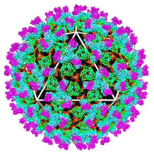 emd_5147_1.jpg emd_5147_1.jpg | 190.1 KB | ||
| Masks | emd_5147_msk_1.mapemd_5147_msk_2.map | 11.4 MB 4.4 MB | Mask map | |
| Archive directory |  http://ftp.pdbj.org/pub/emdb/structures/EMD-5147ftp://ftp.pdbj.org/pub/emdb/structures/EMD-5147 http://ftp.pdbj.org/pub/emdb/structures/EMD-5147ftp://ftp.pdbj.org/pub/emdb/structures/EMD-5147 | HTTPS FTP |
-Related structure data
| Related structure data |  3iykMC M: atomic model generated by this map C: citing same article ( |
|---|---|
| Similar structure data |


-Links
| EMDB pages | EMDB (EBI/PDBe) / EMDataResource |
|---|---|
| Related items in Molecule of the Month |

-Map
| File | Download / File: emd_5147.map.gz / Format: CCP4 / Size: 465.7 MB / Type: IMAGE STORED AS FLOATING POINT NUMBER (4 BYTES) | ||||||||||||||||||||||||||||||||||||||||||||||||||||||||||||||||||||
|---|---|---|---|---|---|---|---|---|---|---|---|---|---|---|---|---|---|---|---|---|---|---|---|---|---|---|---|---|---|---|---|---|---|---|---|---|---|---|---|---|---|---|---|---|---|---|---|---|---|---|---|---|---|---|---|---|---|---|---|---|---|---|---|---|---|---|---|---|---|
| Annotation | BTV map | ||||||||||||||||||||||||||||||||||||||||||||||||||||||||||||||||||||
| Projections & slices | Image control
Images are generated by Spider. | ||||||||||||||||||||||||||||||||||||||||||||||||||||||||||||||||||||
| Voxel size | X=Y=Z: 1.88 Å | ||||||||||||||||||||||||||||||||||||||||||||||||||||||||||||||||||||
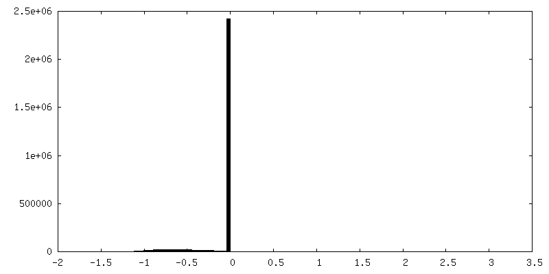
| Density |
| ||||||||||||||||||||||||||||||||||||||||||||||||||||||||||||||||||||
| Symmetry | Space group: 1 | ||||||||||||||||||||||||||||||||||||||||||||||||||||||||||||||||||||
| Details | EMDB XML:
CCP4 map header:
| ||||||||||||||||||||||||||||||||||||||||||||||||||||||||||||||||||||
 Z (Sec.)
 Y (Row.)
 X (Col.)


-Supplemental data
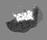
-Segmentation: BTV VP2
| Annotation | BTV VP2 | ||||||||||||
|---|---|---|---|---|---|---|---|---|---|---|---|---|---|
| File | emd_5147_msk_1.map | ||||||||||||
| Projections & Slices |
| ||||||||||||
| Density Histograms |


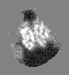
-Segmentation: averaged VP5
| Annotation | averaged VP5 | ||||||||||||
|---|---|---|---|---|---|---|---|---|---|---|---|---|---|
| File | emd_5147_msk_2.map | ||||||||||||
| Projections & Slices |
| ||||||||||||
| Density Histograms |


- Sample components
-Entire : Bluetongue Virus
| Entire | Name: Bluetongue Virus |
|---|---|
| Components |
|
-Supramolecule #1000: Bluetongue Virus
| Supramolecule | Name: Bluetongue Virus / type: sample / ID: 1000 / Details: The sample was monodisperse in ice / Oligomeric state: Icosahedron / Number unique components: 4 |
|---|
-Supramolecule #1: Bluetongue virus
| Supramolecule | Name: Bluetongue virus / type: virus / ID: 1 / Name.synonym: Bluetongue Virus / NCBI-ID: 40051 / Sci species name: Bluetongue virus / Database: NCBI / Virus type: VIRION / Virus isolate: STRAIN / Virus enveloped: No / Virus empty: No / Syn species name: Bluetongue Virus |
|---|---|
| Host (natural) | Organism: livestock (unknown) / synonym: VERTEBRATES |
-Experimental details
-Structure determination
| Method | negative staining, cryo EM |
|---|---|
 Processing Processing | single particle reconstruction |
| Aggregation state | particle |
-Sample preparation
| Buffer | pH: 8 / Details: PBS |
|---|---|
| Staining | Type: NEGATIVE / Details: -175C |
| Grid | Details: 400 mesh Lacey carbon film |
| Vitrification | Cryogen name: ETHANE / Chamber temperature: 90 K / Instrument: HOMEMADE PLUNGER / Details: Vitrification instrument: manual plunger |
- Electron microscopy
| Microscope | FEI TECNAI F20 |
|---|---|
| Temperature | Average: 90 K |
| Image recording | Category: CCD / Film or detector model: GENERIC CCD / Digitization - Sampling interval: 15 µm / Average electron dose: 25 e/Å2 / Bits/pixel: 16 |
| Electron beam | Acceleration voltage: 200 kV / Electron source:  FIELD EMISSION GUN FIELD EMISSION GUN |
| Electron optics | Calibrated magnification: 79787 / Illumination mode: FLOOD BEAM / Imaging mode: BRIGHT FIELD / Cs: 2 mm / Nominal magnification: 80000 |
| Sample stage | Specimen holder: Gatan 626 / Specimen holder model: GATAN LIQUID NITROGEN |
| Experimental equipment |  Model: Tecnai F20 / Image courtesy: FEI Company |
-Image processing
| CTF correction | Details: Each particle |
|---|---|
| Final reconstruction | Algorithm: OTHER / Resolution.type: BY AUTHOR / Resolution: 7.0 Å / Resolution method: FSC 0.143 CUT-OFF / Software - Name: Frealign IMIRS |
