 Movie
Movie Controller


+ Open data

- Basic information
| Entry |  | |||||||||
|---|---|---|---|---|---|---|---|---|---|---|
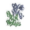
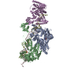
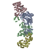
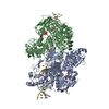
| Title | MtUvrA2 bound to endogenous E. coli DNA at low resolution | |||||||||
 Map data Map data | ||||||||||
 Sample Sample |
| |||||||||
 Keywords Keywords | DNA repair / NER / UVRA / UVRB / UVR / MTB / DNA BINDING PROTEIN | |||||||||
| Biological species |   Mycobacterium tuberculosis (bacteria) / Mycobacterium tuberculosis (bacteria) / | |||||||||
| Method | single particle reconstruction / cryo EM / Resolution: 4.2 Å | |||||||||
 Authors Authors | Genta M / Capelli R / Ferrara G / Rizzi M / Rossi F / Jeruzalmi D / Bolognesi M / Chaves-Sanjuan A / Miggiano R | |||||||||
| Funding support |  Italy, 1 items Italy, 1 items
| |||||||||
 Citation Citation | Journal: Nat Commun / Year: 2025 Title: Mechanistic understanding of UvrA damage detection and lesion hand-off to UvrB in Nucleotide Excision Repair. Authors: Marianna Genta / Giulia Ferrara / Riccardo Capelli / Diego Rondelli / Sarah Sertic / Martino Bolognesi / Menico Rizzi / Franca Rossi / David Jeruzalmi / Antonio Chaves-Sanjuan / Riccardo Miggiano /  Abstract: Nucleotide excision repair (NER) represents one of the major molecular machineries that control chromosome stability in all living species. In Eubacteria, the initial stages of the repair process are ...Nucleotide excision repair (NER) represents one of the major molecular machineries that control chromosome stability in all living species. In Eubacteria, the initial stages of the repair process are carried out by the UvrABC excinuclease complex. Despite the wealth of structural data available, some crucial details of the pathway remain elusive. In this study, we present a structural investigation of the Mycobacterium tuberculosis UvrAUvrB complex and of the UvrA dimer, both in complex with damaged DNA. Our analyses yield insights into the DNA binding mode of UvrA, showing an unexplored conformation of Insertion Domains (IDs), underlying the essential role of these domains in DNA coordination. Furthermore, we observe an interplay between the ID and the UvrB Binding Domain (UBD): after the recognition of the damage, the IDs repositions with the concomitant reorganization of UBD, allowing the formation of the complex between UvrA and UvrB. These events are detected along the formation of the uncharacterized UvrAUvrB-DNA and the UvrAUvrB-DNA complexes which we interpret as hierarchical steps initiating the DNA repair cascade in the NER pathway, resulting in the formation of the pre-incision complex. | |||||||||
| History |
|
- Structure visualization
| Supplemental images |
|---|

- Downloads & links
-EMDB archive
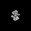
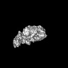
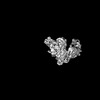
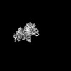
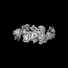
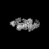
| Map data | emd_51168.map.gz | 120.9 MB |  EMDB map data format EMDB map data format | |
|---|---|---|---|---|
| Header (meta data) | emd-51168-v30.xmlemd-51168.xml | 22.4 KB 22.4 KB | Display Display | EMDB header |
| Images |  emd_51168.png emd_51168.png | 63.6 KB | ||
| Masks | emd_51168_msk_1.map | 244.1 MB | Mask map | |
| Filedesc metadata | emd-51168.cif.gz | 6.2 KB | ||
| Others | emd_51168_additional_1.map.gzemd_51168_half_map_1.map.gzemd_51168_half_map_2.map.gz | 230.1 MB 226.4 MB 226.4 MB | ||
| Archive directory |  http://ftp.pdbj.org/pub/emdb/structures/EMD-51168ftp://ftp.pdbj.org/pub/emdb/structures/EMD-51168 http://ftp.pdbj.org/pub/emdb/structures/EMD-51168ftp://ftp.pdbj.org/pub/emdb/structures/EMD-51168 | HTTPS FTP |
-Related structure data
-Links
| EMDB pages | EMDB (EBI/PDBe) / EMDataResource |
|---|
-Map
| File | Download / File: emd_51168.map.gz / Format: CCP4 / Size: 244.1 MB / Type: IMAGE STORED AS FLOATING POINT NUMBER (4 BYTES) | ||||||||||||||||||||||||||||||||||||
|---|---|---|---|---|---|---|---|---|---|---|---|---|---|---|---|---|---|---|---|---|---|---|---|---|---|---|---|---|---|---|---|---|---|---|---|---|---|
| Projections & slices | Image control
Images are generated by Spider. | ||||||||||||||||||||||||||||||||||||
| Voxel size | X=Y=Z: 0.889 Å | ||||||||||||||||||||||||||||||||||||
| Density |
| ||||||||||||||||||||||||||||||||||||
| Symmetry | Space group: 1 | ||||||||||||||||||||||||||||||||||||
| Details | EMDB XML:
|
 Z (Sec.)
 Y (Row.)
 X (Col.)


-Supplemental data
-Mask #1
| File | emd_51168_msk_1.map | ||||||||||||
|---|---|---|---|---|---|---|---|---|---|---|---|---|---|
| Projections & Slices |
| ||||||||||||
| Density Histograms |


-Additional map: sharp
| File | emd_51168_additional_1.map | ||||||||||||
|---|---|---|---|---|---|---|---|---|---|---|---|---|---|
| Annotation | sharp | ||||||||||||
| Projections & Slices |
| ||||||||||||
| Density Histograms |


-Half map: #1
| File | emd_51168_half_map_1.map | ||||||||||||
|---|---|---|---|---|---|---|---|---|---|---|---|---|---|
| Projections & Slices |
| ||||||||||||
| Density Histograms |


-Half map: #2
| File | emd_51168_half_map_2.map | ||||||||||||
|---|---|---|---|---|---|---|---|---|---|---|---|---|---|
| Projections & Slices |
| ||||||||||||
| Density Histograms |


- Sample components
-Entire : MtUvrA2-DNA complex
| Entire | Name: MtUvrA2-DNA complex |
|---|---|
| Components |
|
-Supramolecule #1: MtUvrA2-DNA complex
| Supramolecule | Name: MtUvrA2-DNA complex / type: complex / ID: 1 / Parent: 0 / Macromolecule list: all / Details: MtUvrA2 in complex with DNA |
|---|---|
| Source (natural) | Organism: Mycobacterium tuberculosis (bacteria) |
| Molecular weight | Theoretical: 220 KDa |
-Macromolecule #1: MtUvrA
| Macromolecule | Name: MtUvrA / type: protein_or_peptide / ID: 1 / Enantiomer: LEVO |
|---|---|
| Source (natural) | Organism: Mycobacterium tuberculosis (bacteria) |
| Recombinant expression | Organism: |
| Sequence | String: MGHHHHHHHH HHSSGHIEGR HMADRLIVKG AREHNLRSVD LDLPRDALIV FTGLSGSGKS SLAFDTIFAE GQRRYVESLS AYARQFLGQM DKPDVDFIEG LSPAVSIDQK STNRNPRSTV GTITEVYDYL RLLYARAGTP HCPTCGERVA RQTPQQIVDQ VLAMPEGTRF ...String: MGHHHHHHHH HHSSGHIEGR HMADRLIVKG AREHNLRSVD LDLPRDALIV FTGLSGSGKS SLAFDTIFAE GQRRYVESLS AYARQFLGQM DKPDVDFIEG LSPAVSIDQK STNRNPRSTV GTITEVYDYL RLLYARAGTP HCPTCGERVA RQTPQQIVDQ VLAMPEGTRF LVLAPVVRTR KGEFADLFDK LNAQGYSRVR VDGVVHPLTD PPKLKKQEKH DIEVVVDRLT VKAAAKRRLT DSVETALNLA DGIVVLEFVD HELGAPHREQ RFSEKLACPN GHALAVDDLE PRSFSFNSPY GACPECSGLG IRKEVDPELV VPDPDRTLAQ GAVAPWSNGH TAEYFTRMMA GLGEALGFDV DTPWRKLPAK ARKAILEGAD EQVHVRYRNR YGRTRSYYAD FEGVLAFLQR KMSQTESEQM KERYEGFMRD VPCPVCAGTR LKPEILAVTL AGESKGEHGA KSIAEVCELS IADCADFLNA LTLGPREQAI AGQVLKEIRS RLGFLLDVGL EYLSLSRAAA TLSGGEAQRI RLATQIGSGL VGVLYVLDEP SIGLHQRDNR RLIETLTRLR DLGNTLIVVE HDEDTIEHAD WIVDIGPGAG EHGGRIVHSG PYDELLRNKD SITGAYLSGR ESIEIPAIRR SVDPRRQLTV VGAREHNLRG IDVSFPLGVL TSVTGVSGSG KSTLVNDILA AVLANRLNGA RQVPGRHTRV TGLDYLDKLV RVDQSPIGRT PRSNPATYTG VFDKIRTLFA ATTEAKVRGY QPGRFSFNVK GGRCEACTGD GTIKIEMNFL PDVYVPCEVC QGARYNRETL EVHYKGKTVS EVLDMSIEEA AEFFEPIAGV HRYLRTLVDV GLGYVRLGQP APTLSGGEAQ RVKLASELQK RSTGRTVYIL DEPTTGLHFD DIRKLLNVIN GLVDKGNTVI VIEHNLDVIK TSDWIIDLGP EGGAGGGTVV AQGTPEDVAA VPASYTGKFL AEVVGGGASA ATSRSNRRRN VSA |
-Macromolecule #2: Endogenous E. coli DNA
| Macromolecule | Name: Endogenous E. coli DNA / type: dna / ID: 2 / Classification: DNA |
|---|---|
| Source (natural) | Organism: |
| Sequence | String: CACATGAAAA AAAAAATGAA AATGATTTCT GA |
-Experimental details
-Structure determination
| Method | cryo EM |
|---|---|
 Processing Processing | single particle reconstruction |
| Aggregation state | particle |
-Sample preparation
| Concentration | 0.5 mg/mL | |||||||||
|---|---|---|---|---|---|---|---|---|---|---|
| Buffer | pH: 8 Component:
| |||||||||
| Grid | Model: Quantifoil R1.2/1.3 / Material: COPPER / Mesh: 300 / Support film - Material: CARBON / Support film - topology: HOLEY / Pretreatment - Type: GLOW DISCHARGE / Pretreatment - Time: 30 sec. / Pretreatment - Atmosphere: AIR / Details: 30mA | |||||||||
| Vitrification | Cryogen name: ETHANE / Chamber humidity: 100 % / Chamber temperature: 277 K / Instrument: FEI VITROBOT MARK IV |
- Electron microscopy
| Microscope | FEI TALOS ARCTICA |
|---|---|
| Image recording | Film or detector model: FEI FALCON III (4k x 4k) / Detector mode: COUNTING / Number grids imaged: 1 / Number real images: 3642 / Average electron dose: 40.0 e/Å2 |
| Electron beam | Acceleration voltage: 200 kV / Electron source:  FIELD EMISSION GUN FIELD EMISSION GUN |
| Electron optics | Illumination mode: FLOOD BEAM / Imaging mode: BRIGHT FIELD / Cs: 2.7 mm / Nominal defocus max: 2.5 µm / Nominal defocus min: 0.8 µm / Nominal magnification: 120000 |
| Sample stage | Specimen holder model: FEI TITAN KRIOS AUTOGRID HOLDER / Cooling holder cryogen: NITROGEN |
| Experimental equipment |  Model: Talos Arctica / Image courtesy: FEI Company |
