 Movie
Movie Controller

[English] 日本語
 Yorodumi
- EMDB-50845: Real space helical reconstruction of cofilin actin in the microtu... -
+ Open data

- Basic information
| Entry |  | |||||||||||||||||||||||||||||||||||||||
|---|---|---|---|---|---|---|---|---|---|---|---|---|---|---|---|---|---|---|---|---|---|---|---|---|---|---|---|---|---|---|---|---|---|---|---|---|---|---|---|---|
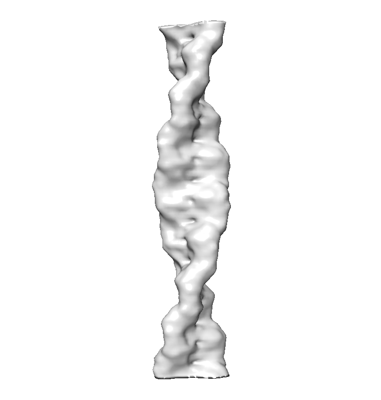
| Title | Real space helical reconstruction of cofilin actin in the microtubule lumen of HAP1 cells | |||||||||||||||||||||||||||||||||||||||
 Map data Map data | Real space helical reconstruction of cofilin actin in the microtubule lumen of HAP1 cells | |||||||||||||||||||||||||||||||||||||||
 Sample Sample |
| |||||||||||||||||||||||||||||||||||||||
 Keywords Keywords | cofilin / actin / filament / CYTOSOLIC PROTEIN | |||||||||||||||||||||||||||||||||||||||
| Biological species |  Homo sapiens (human) Homo sapiens (human) | |||||||||||||||||||||||||||||||||||||||
| Method | helical reconstruction / cryo EM / Resolution: 20.0 Å | |||||||||||||||||||||||||||||||||||||||
 Authors Authors | Tsuji C / Bradshaw M / Paul DM / Dodding MP | |||||||||||||||||||||||||||||||||||||||
| Funding support |  United Kingdom, 12 items United Kingdom, 12 items
| |||||||||||||||||||||||||||||||||||||||
 Citation Citation | Journal: Nat Commun / Year: 2024 Title: CryoET reveals actin filaments within platelet microtubules. Authors: Chisato Tsuji / Marston Bradshaw / Megan F Allen / Molly L Jackson / Judith Mantell / Ufuk Borucu / Alastair W Poole / Paul Verkade / Ingeborg Hers / Danielle M Paul / Mark P Dodding / Abstract: Crosstalk between the actin and microtubule cytoskeletons is important for many cellular processes. Recent studies have shown that microtubules and F-actin can assemble to form a composite structure ...Crosstalk between the actin and microtubule cytoskeletons is important for many cellular processes. Recent studies have shown that microtubules and F-actin can assemble to form a composite structure where F-actin occupies the microtubule lumen. Whether these cytoskeletal hybrids exist in physiological settings and how they are formed is unclear. Here, we show that the short-crossover Class I actin filament previously identified inside microtubules in human HAP1 cells is cofilin-bound F-actin. Lumenal F-actin can be reconstituted in vitro, but cofilin is not essential. Moreover, actin filaments with both cofilin-bound and canonical morphologies reside within human platelet microtubules under physiological conditions. We propose that stress placed upon the microtubule network during motor-driven microtubule looping and sliding may facilitate the incorporation of actin into microtubules. | |||||||||||||||||||||||||||||||||||||||
| History |
|
- Structure visualization
| Supplemental images |
|---|
- Downloads & links
-EMDB archive
| Map data | emd_50845.map.gz | 7.6 MB |  EMDB map data format EMDB map data format | |
|---|---|---|---|---|
| Header (meta data) | emd-50845-v30.xmlemd-50845.xml | 11.3 KB 11.3 KB | Display Display | EMDB header |
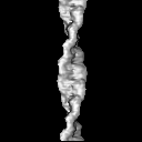
| Images |  emd_50845.png emd_50845.png | 26.7 KB | ||
| Filedesc metadata | emd-50845.cif.gz | 4.1 KB | ||
| Archive directory |  http://ftp.pdbj.org/pub/emdb/structures/EMD-50845ftp://ftp.pdbj.org/pub/emdb/structures/EMD-50845 http://ftp.pdbj.org/pub/emdb/structures/EMD-50845ftp://ftp.pdbj.org/pub/emdb/structures/EMD-50845 | HTTPS FTP |
-Related structure data
-Links
| EMDB pages | EMDB (EBI/PDBe) / EMDataResource |
|---|
-Map
| File | Download / File: emd_50845.map.gz / Format: CCP4 / Size: 8 MB / Type: IMAGE STORED AS FLOATING POINT NUMBER (4 BYTES) | ||||||||||||||||||||||||||||||||||||
|---|---|---|---|---|---|---|---|---|---|---|---|---|---|---|---|---|---|---|---|---|---|---|---|---|---|---|---|---|---|---|---|---|---|---|---|---|---|
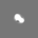
| Annotation | Real space helical reconstruction of cofilin actin in the microtubule lumen of HAP1 cells | ||||||||||||||||||||||||||||||||||||
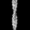
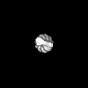
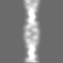
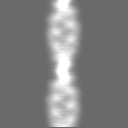
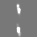
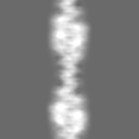
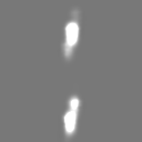
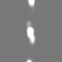
| Projections & slices | Image control
Images are generated by Spider. | ||||||||||||||||||||||||||||||||||||
| Voxel size | X=Y=Z: 3.75 Å | ||||||||||||||||||||||||||||||||||||
| Density |
| ||||||||||||||||||||||||||||||||||||
| Symmetry | Space group: 1 | ||||||||||||||||||||||||||||||||||||
| Details | EMDB XML:
|
 Z (Sec.)
 Y (Row.)
 X (Col.)


-Supplemental data
- Sample components
-Entire : Cofilin bound actin structure found in the lumen of microtubules ...
| Entire | Name: Cofilin bound actin structure found in the lumen of microtubules in HAP1 cells |
|---|---|
| Components |
|
-Supramolecule #1: Cofilin bound actin structure found in the lumen of microtubules ...
| Supramolecule | Name: Cofilin bound actin structure found in the lumen of microtubules in HAP1 cells type: complex / ID: 1 / Parent: 0 |
|---|---|
| Source (natural) | Organism: Homo sapiens (human) / Strain: HAP1 cells |
-Experimental details
-Structure determination
| Method | cryo EM |
|---|---|
 Processing Processing | helical reconstruction |
| Aggregation state | filament |
-Sample preparation
| Buffer | pH: 6.8 |
|---|---|
| Vitrification | Cryogen name: ETHANE / Instrument: LEICA EM GP |
- Electron microscopy
| Microscope | TFS KRIOS |
|---|---|
| Image recording | Film or detector model: TFS FALCON 4i (4k x 4k) / Average electron dose: 2.0 e/Å2 |
| Electron beam | Acceleration voltage: 300 kV / Electron source:  FIELD EMISSION GUN FIELD EMISSION GUN |
| Electron optics | Illumination mode: FLOOD BEAM / Imaging mode: BRIGHT FIELD / Cs: 2.7 mm / Nominal defocus max: 7.0 µm / Nominal defocus min: 3.0 µm / Nominal magnification: 63000 |
| Experimental equipment |  Model: Titan Krios / Image courtesy: FEI Company |
-Image processing
| Final reconstruction | Applied symmetry - Helical parameters - Δz: 27.5 Å Applied symmetry - Helical parameters - Δ&Phi: 162 ° Applied symmetry - Helical parameters - Axial symmetry: C1 (asymmetric) Resolution.type: BY AUTHOR / Resolution: 20.0 Å / Resolution method: OTHER Details: A sub-volume containing the filament was extracted from a tomogram, which was then 2D projected. The projected image was used for real space helical reconstruction to create the map. ...Details: A sub-volume containing the filament was extracted from a tomogram, which was then 2D projected. The projected image was used for real space helical reconstruction to create the map. Resolution was estimated compared to molmap generation of PDB:3J0S at 20 angstroms in Chimera and correlation score of 0.96 was obtained. Number images used: 1 |
|---|---|
| Startup model | Type of model: NONE |
| Final angle assignment | Type: NOT APPLICABLE |
