 Movie
Movie Controller

[English] 日本語
 Yorodumi
- EMDB-50838: Cryo-EM structure of the type 1 pilus assembly platform as part o... -
+ Open data

- Basic information
| Entry |  | |||||||||
|---|---|---|---|---|---|---|---|---|---|---|
| Title | Cryo-EM structure of the type 1 pilus assembly platform as part of the FimA-bound chaperone-usher pilus complex (Local refinement including FimD, FimC, FimAn, FimAn-1 and FimAn-2) | |||||||||
 Map data Map data | Map of the type 1 pilus assembly platform as part of the FimA-bound chaperone-usher pilus complex - Local refinement including FimD, FimC, FimAn, FimAn-1 and FimAn-2 (sharpened) | |||||||||
 Sample Sample |
| |||||||||
 Keywords Keywords | chaperone / usher / pilus / rod / MEMBRANE PROTEIN | |||||||||
| Function / homology |  Function and homology information Function and homology informationfimbrial usher porin activity / pilus assembly / cell adhesion involved in single-species biofilm formation / pilus / protein folding chaperone / cell outer membrane / cell wall organization / outer membrane-bounded periplasmic space / protein folding / cell adhesion / identical protein binding Similarity search - Function | |||||||||
| Biological species |  | |||||||||
| Method | single particle reconstruction / cryo EM / Resolution: 3.6 Å | |||||||||
 Authors Authors | Bachmann P / Afanasyev P / Boehringer D / Glockshuber R | |||||||||
| Funding support |  Switzerland, 1 items Switzerland, 1 items
| |||||||||
 Citation Citation | Journal: To Be Published Title: Cryo-EM structure of the type 1 pilus assembly platform as part of the FimA-bound chaperone-usher pilus complex (Local refinement including FimD, FimC, FimAn, FimAn-1 and FimAn-2) Authors: Bachmann P / Afanasyev P / Boehringer D / Glockshuber R | |||||||||
| History |
|
- Structure visualization
| Supplemental images |
|---|

- Downloads & links
-EMDB archive
| Map data | emd_50838.map.gz | 59.4 MB | EMDB map data format | |
|---|---|---|---|---|
| Header (meta data) | emd-50838-v30.xmlemd-50838.xml | 22.1 KB 22.1 KB | Display Display | EMDB header |
| FSC (resolution estimation) | emd_50838_fsc.xml | 9.6 KB | Display | FSC data file |
| Images |  emd_50838.png emd_50838.png | 106.8 KB | ||
| Filedesc metadata | emd-50838.cif.gz | 6.5 KB | ||
| Others | emd_50838_additional_1.map.gzemd_50838_half_map_1.map.gzemd_50838_half_map_2.map.gz | 32.3 MB 59.4 MB 59.4 MB | ||
| Archive directory |  http://ftp.pdbj.org/pub/emdb/structures/EMD-50838ftp://ftp.pdbj.org/pub/emdb/structures/EMD-50838 http://ftp.pdbj.org/pub/emdb/structures/EMD-50838ftp://ftp.pdbj.org/pub/emdb/structures/EMD-50838 | HTTPS FTP |
-Related structure data
| Related structure data |  9fwzMC M: atomic model generated by this map C: citing same article ( |
|---|---|
| Similar structure data |
-Links
| EMDB pages | EMDB (EBI/PDBe) / EMDataResource |
|---|
-Map
| File | Download / File: emd_50838.map.gz / Format: CCP4 / Size: 64 MB / Type: IMAGE STORED AS FLOATING POINT NUMBER (4 BYTES) | ||||||||||||||||||||||||||||||||||||
|---|---|---|---|---|---|---|---|---|---|---|---|---|---|---|---|---|---|---|---|---|---|---|---|---|---|---|---|---|---|---|---|---|---|---|---|---|---|
| Annotation | Map of the type 1 pilus assembly platform as part of the FimA-bound chaperone-usher pilus complex - Local refinement including FimD, FimC, FimAn, FimAn-1 and FimAn-2 (sharpened) | ||||||||||||||||||||||||||||||||||||
| Projections & slices | Image control
Images are generated by Spider. | ||||||||||||||||||||||||||||||||||||
| Voxel size | X=Y=Z: 1.296 Å | ||||||||||||||||||||||||||||||||||||
| Density |
| ||||||||||||||||||||||||||||||||||||
| Symmetry | Space group: 1 | ||||||||||||||||||||||||||||||||||||
| Details | EMDB XML:
|
 Z (Sec.)
 Y (Row.)
 X (Col.)


-Supplemental data
-Additional map: Map of the type 1 pilus assembly platform...
| File | emd_50838_additional_1.map | ||||||||||||
|---|---|---|---|---|---|---|---|---|---|---|---|---|---|
| Annotation | Map of the type 1 pilus assembly platform as part of the FimA-bound chaperone-usher pilus complex - Local refinement including FimD, FimC, FimAn, FimAn-1 and FimAn-2 (unsharpened) | ||||||||||||
| Projections & Slices |
| ||||||||||||
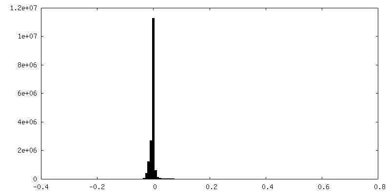
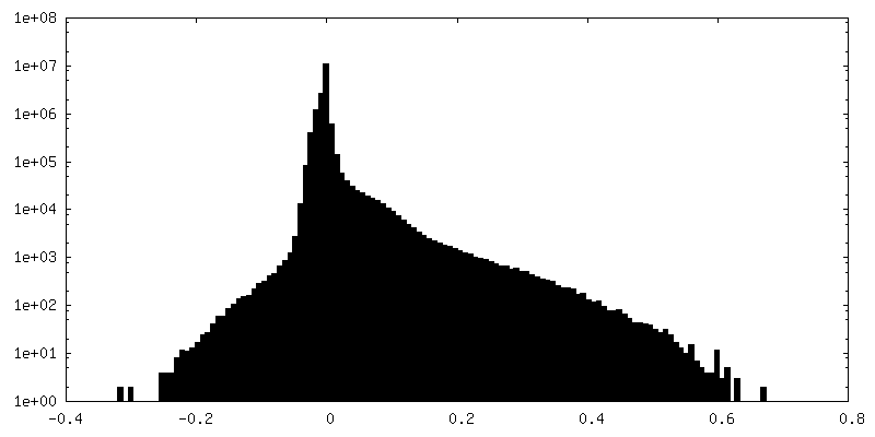
| Density Histograms |


-Half map: Half-map A of the type 1 pilus assembly...
| File | emd_50838_half_map_1.map | ||||||||||||
|---|---|---|---|---|---|---|---|---|---|---|---|---|---|
| Annotation | Half-map A of the type 1 pilus assembly platform as part of the FimA-bound chaperone-usher pilus complex - Local refinement including FimD, FimC, FimAn, FimAn-1 and FimAn-2 | ||||||||||||
| Projections & Slices |
| ||||||||||||
| Density Histograms |


-Half map: Half-map B of the type 1 pilus assembly...
| File | emd_50838_half_map_2.map | ||||||||||||
|---|---|---|---|---|---|---|---|---|---|---|---|---|---|
| Annotation | Half-map B of the type 1 pilus assembly platform as part of the FimA-bound chaperone-usher pilus complex - Local refinement including FimD, FimC, FimAn, FimAn-1 and FimAn-2 | ||||||||||||
| Projections & Slices |
| ||||||||||||
| Density Histograms |


- Sample components
-Entire : FimDHGFAnC complex
| Entire | Name: FimDHGFAnC complex |
|---|---|
| Components |
|
-Supramolecule #1: FimDHGFAnC complex
| Supramolecule | Name: FimDHGFAnC complex / type: complex / ID: 1 / Parent: 0 / Macromolecule list: all |
|---|---|
| Source (natural) | Organism: |
-Macromolecule #1: Outer membrane usher protein FimD
| Macromolecule | Name: Outer membrane usher protein FimD / type: protein_or_peptide / ID: 1 / Number of copies: 1 / Enantiomer: LEVO |
|---|---|
| Source (natural) | Organism: |
| Molecular weight | Theoretical: 93.092805 KDa |
| Recombinant expression | Organism: |
| Sequence | String: DLYFNPRFLA DDPQAVADLS RFENGQELPP GTYRVDIYLN NGYMATRDVT FNTGDSEQGI VPCLTRAQLA SMGLNTASVA GMNLLADDA CVPLTTMVQD ATAHLDVGQQ RLNLTIPQAF MSNRARGYIP PELWDPGINA GLLNYNFSGN SVQNRIGGNS H YAYLNLQS ...String: DLYFNPRFLA DDPQAVADLS RFENGQELPP GTYRVDIYLN NGYMATRDVT FNTGDSEQGI VPCLTRAQLA SMGLNTASVA GMNLLADDA CVPLTTMVQD ATAHLDVGQQ RLNLTIPQAF MSNRARGYIP PELWDPGINA GLLNYNFSGN SVQNRIGGNS H YAYLNLQS GLNIGAWRLR DNTTWSYNSS DRSSGSKNKW QHINTWLERD IIPLRSRLTL GDGYTQGDIF DGINFRGAQL AS DDNMLPD SQRGFAPVIH GIARGTAQVT IKQNGYDIYN STVPPGPFTI NDIYAAGNSG DLQVTIKEAD GSTQIFTVPY SSV PLLQRE GHTRYSITAG EYRSGNAQQE KPRFFQSTLL HGLPAGWTIY GGTQLADRYR AFNFGIGKNM GALGALSVDM TQAN STLPD DSQHDGQSVR FLYNKSLNES GTNIQLVGYR YSTSGYFNFA DTTYSRMNGY NIETQDGVIQ VKPKFTDYYN LAYNK RGKL QLTVTQQLGR TSTLYLSGSH QTYWGTSNVD EQFQAGLNTA FEDINWTLSY SLTKNAWQKG RDQMLALNVN IPFSHW LRS DSKSQWRHAS ASYSMSHDLN GRMTNLAGVY GTLLEDNNLS YSVQTGYAGG GDGNSGSTGY ATLNYRGGYG NANIGYS HS DDIKQLYYGV SGGVLAHANG VTLGQPLNDT VVLVKAPGAK DAKVENQTGV RTDWRGYAVL PYATEYRENR VALDTNTL A DNVDLDNAVA NVVPTRGAIV RAEFKARVGI KLLMTLTHNN KPLPFGAMVT SESSQSSGIV ADNGQVYLSG MPLAGKVQV KWGEEENAHC VANYQLPPES QQQLLTQLSA ECRLVPRGSW SHPQFEK UniProtKB: Outer membrane usher protein FimD |
-Macromolecule #2: Chaperone protein FimC
| Macromolecule | Name: Chaperone protein FimC / type: protein_or_peptide / ID: 2 / Number of copies: 1 / Enantiomer: LEVO |
|---|---|
| Source (natural) | Organism: |
| Molecular weight | Theoretical: 22.885229 KDa |
| Recombinant expression | Organism: |
| Sequence | String: MGVALGATRV IYPAGQKQEQ LAVTNNDENS TYLIQSWVEN ADGVKDGRFI VTPPLFAMKG KKENTLRILD ATNNQLPQDR ESLFWMNVK AIPSMDKSKL TENTLQLAII SRIKLYYRPA KLALPPDQAA EKLRFRRSAN SLTLINPTPY YLTVTELNAG T RVLENALV ...String: MGVALGATRV IYPAGQKQEQ LAVTNNDENS TYLIQSWVEN ADGVKDGRFI VTPPLFAMKG KKENTLRILD ATNNQLPQDR ESLFWMNVK AIPSMDKSKL TENTLQLAII SRIKLYYRPA KLALPPDQAA EKLRFRRSAN SLTLINPTPY YLTVTELNAG T RVLENALV PPMGESTVKL PSDAGSNITY RTINDYGALT PKMTGVME UniProtKB: Chaperone protein FimC |
-Macromolecule #3: Type-1 fimbrial protein, A chain
| Macromolecule | Name: Type-1 fimbrial protein, A chain / type: protein_or_peptide / ID: 3 / Number of copies: 3 / Enantiomer: LEVO |
|---|---|
| Source (natural) | Organism: |
| Molecular weight | Theoretical: 15.96644 KDa |
| Recombinant expression | Organism: |
| Sequence | String: MAATTVNGGT VHFKGEVVNA ACAVDAGSVD QTVQLGQVRT ASLAQEGATS SAVGFNIQLN DCDTNVASKA AVAFLGTAID AGHTNVLAL QSSAAGSATN VGVQILDRTG AALTLDGATF SSETTLNNGT NTIPFQARYF ATGAATPGAA NADATFKVQY Q UniProtKB: Type-1 fimbrial protein, A chain |
-Experimental details
-Structure determination
| Method | cryo EM |
|---|---|
 Processing Processing | single particle reconstruction |
| Aggregation state | particle |
-Sample preparation
| Buffer | pH: 8 |
|---|---|
| Grid | Model: Quantifoil R2/2 / Material: COPPER / Mesh: 300 |
| Vitrification | Cryogen name: ETHANE-PROPANE / Chamber humidity: 100 % / Chamber temperature: 277 K / Instrument: FEI VITROBOT MARK IV |
- Electron microscopy
| Microscope | FEI TITAN KRIOS |
|---|---|
| Image recording | Film or detector model: GATAN K3 BIOQUANTUM (6k x 4k) / Average exposure time: 1.1 sec. / Average electron dose: 64.0 e/Å2 |
| Electron beam | Acceleration voltage: 300 kV / Electron source:  FIELD EMISSION GUN FIELD EMISSION GUN |
| Electron optics | C2 aperture diameter: 100.0 µm / Illumination mode: FLOOD BEAM / Imaging mode: BRIGHT FIELD / Cs: 2.7 mm / Nominal defocus max: 2.8000000000000003 µm / Nominal defocus min: 1.0 µm / Nominal magnification: 130000 |
| Sample stage | Specimen holder model: FEI TITAN KRIOS AUTOGRID HOLDER / Cooling holder cryogen: NITROGEN |
| Experimental equipment |  Model: Titan Krios / Image courtesy: FEI Company |

