Biotechnology and Biological Sciences Research Council (BBSRC)
United Kingdom
Citation
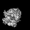
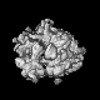
Journal: Nucleic Acids Res / Year: 2025 Title: Translational activity of 80S monosomes varies dramatically across different tissues. Authors: Albert Blandy / Tayah Hopes / Elton J R Vasconcelos / Amy Turner / Bulat Fatkhullin / Michaela Agapiou / Juan Fontana / Julie L Aspden / Abstract: Translational regulation at the stage of initiation can impact the number of ribosomes translating each mRNA molecule. However, the translational activity of single 80S ribosomes (monosomes) on mRNA ...Translational regulation at the stage of initiation can impact the number of ribosomes translating each mRNA molecule. However, the translational activity of single 80S ribosomes (monosomes) on mRNA is less well understood, even though these 80S monosomes represent the dominant ribosomal complexes in vivo. Here, we used cryo-EM to determine the translational activity of 80S monosomes across different tissues in Drosophila melanogaster. We discovered that while head and embryo 80S monosomes are highly translationally active, testis and ovary 80S monosomes are translationally inactive. RNA-Seq analysis of head monosome- and polysome-translated mRNAs, revealed that head 80S monosomes preferentially translate mRNAs with TOP motifs, short 5'-UTRs, short ORFs and are enriched for the presence of uORFs. Overall, these findings highlight that regulation of translation initiation and protein synthesis is mostly performed by monosomes in head and embryo, while polysomes are the main source of protein production in testis and ovary.
In the structure databanks used in Yorodumi, some data are registered as the other names, "COVID-19 virus" and "2019-nCoV". Here are the details of the virus and the list of structure data.
Jan 31, 2019. EMDB accession codes are about to change! (news from PDBe EMDB page)
EMDB accession codes are about to change! (news from PDBe EMDB page)
The allocation of 4 digits for EMDB accession codes will soon come to an end. Whilst these codes will remain in use, new EMDB accession codes will include an additional digit and will expand incrementally as the available range of codes is exhausted. The current 4-digit format prefixed with “EMD-” (i.e. EMD-XXXX) will advance to a 5-digit format (i.e. EMD-XXXXX), and so on. It is currently estimated that the 4-digit codes will be depleted around Spring 2019, at which point the 5-digit format will come into force.
The EM Navigator/Yorodumi systems omit the EMD- prefix.
Related info.:Q: What is EMD? / ID/Accession-code notation in Yorodumi/EM Navigator
Yorodumi is a browser for structure data from EMDB, PDB, SASBDB, etc.
This page is also the successor to EM Navigator detail page, and also detail information page/front-end page for Omokage search.
The word "yorodu" (or yorozu) is an old Japanese word meaning "ten thousand". "mi" (miru) is to see.
Related info.:EMDB / PDB / SASBDB / Comparison of 3 databanks / Yorodumi Search / Aug 31, 2016. New EM Navigator & Yorodumi / Yorodumi Papers / Jmol/JSmol / Function and homology information / Changes in new EM Navigator and Yorodumi
 Movie
Movie Controller


 Open data
 Basic information
 Map data
 Sample
 Keywords
 Authors
 United Kingdom, 1 items
  Citation
 Structure visualization
 Downloads & links
 EMDB map data format
 emd_50765.png
 http://ftp.pdbj.org/pub/emdb/structures/EMD-50765


 Z (Sec.)
 Y (Row.)
 X (Col.)


 Sample components
 Processing
 Electron microscopy
 FIELD EMISSION GUN
