 Movie
Movie Controller

[English] 日本語
 Yorodumi
- EMDB-50744: Closed conformation of the pentameric ligand-gated ion channel, D... -
+ Open data

- Basic information
| Entry |  | |||||||||
|---|---|---|---|---|---|---|---|---|---|---|
| Title | Closed conformation of the pentameric ligand-gated ion channel, DeCLIC at pH 5 with 10 mM Ca2+ | |||||||||
 Map data Map data | ||||||||||
 Sample Sample |
| |||||||||
 Keywords Keywords | Pentameric ligand-gated ion channel / bacterial ion channel / MEMBRANE PROTEIN | |||||||||
| Function / homology |  Function and homology information Function and homology informationextracellular ligand-gated monoatomic ion channel activity / transmembrane signaling receptor activity / membrane / metal ion binding Similarity search - Function | |||||||||
| Biological species |  Desulfacinum sp. (bacteria) / Desulfofustis sp. PB-SRB1 (bacteria) Desulfacinum sp. (bacteria) / Desulfofustis sp. PB-SRB1 (bacteria) | |||||||||
| Method | single particle reconstruction / cryo EM / Resolution: 3.1 Å | |||||||||
 Authors Authors | Rovsnik U / Anden O / Lycksell M / Delarue M / Howard RJ / Lindahl E | |||||||||
| Funding support |  Sweden, 2 items Sweden, 2 items
| |||||||||
 Citation Citation | Journal: To Be Published Title: Structural characterization of pH-modulated closed and open states in a pentameric ligand-gated ion channel Authors: Rovsnik U / Anden O / Lycksell M / Delarue M / Howard RJ / Lindahl E | |||||||||
| History |
|
- Structure visualization
| Supplemental images |
|---|

- Downloads & links
-EMDB archive
| Map data | emd_50744.map.gz | 58.9 MB | EMDB map data format | |
|---|---|---|---|---|
| Header (meta data) | emd-50744-v30.xmlemd-50744.xml | 19 KB 19 KB | Display Display | EMDB header |
| FSC (resolution estimation) | emd_50744_fsc.xml | 9.1 KB | Display | FSC data file |
| Images |  emd_50744.png emd_50744.png | 148.6 KB | ||
| Masks | emd_50744_msk_1.map | 64 MB | Mask map | |
| Filedesc metadata | emd-50744.cif.gz | 6.5 KB | ||
| Others | emd_50744_half_map_1.map.gzemd_50744_half_map_2.map.gz | 49.7 MB 50 MB | ||
| Archive directory |  http://ftp.pdbj.org/pub/emdb/structures/EMD-50744ftp://ftp.pdbj.org/pub/emdb/structures/EMD-50744 http://ftp.pdbj.org/pub/emdb/structures/EMD-50744ftp://ftp.pdbj.org/pub/emdb/structures/EMD-50744 | HTTPS FTP |
-Related structure data
| Related structure data | 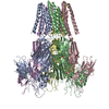 9ftgMC 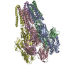 9fthC 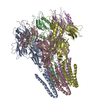 9ftiC 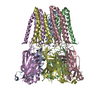 9ftjC M: atomic model generated by this map C: citing same article ( |
|---|---|
| Similar structure data |
-Links
| EMDB pages | EMDB (EBI/PDBe) / EMDataResource |
|---|---|
| Related items in Molecule of the Month |


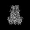
-Map
| File | Download / File: emd_50744.map.gz / Format: CCP4 / Size: 64 MB / Type: IMAGE STORED AS FLOATING POINT NUMBER (4 BYTES) | ||||||||||||||||||||||||||||||||||||
|---|---|---|---|---|---|---|---|---|---|---|---|---|---|---|---|---|---|---|---|---|---|---|---|---|---|---|---|---|---|---|---|---|---|---|---|---|---|
| Projections & slices | Image control
Images are generated by Spider. | ||||||||||||||||||||||||||||||||||||
| Voxel size | X=Y=Z: 0.8617 Å | ||||||||||||||||||||||||||||||||||||
| Density |
| ||||||||||||||||||||||||||||||||||||
| Symmetry | Space group: 1 | ||||||||||||||||||||||||||||||||||||
| Details | EMDB XML:
|
 Z (Sec.)
 Y (Row.)
 X (Col.)


-Supplemental data
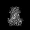
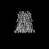
-Mask #1
| File | emd_50744_msk_1.map | ||||||||||||
|---|---|---|---|---|---|---|---|---|---|---|---|---|---|
| Projections & Slices |
| ||||||||||||
| Density Histograms |


- Sample components
-Entire : Pentameric ligan-gated ion channel DeCLIC
| Entire | Name: Pentameric ligan-gated ion channel DeCLIC |
|---|---|
| Components |
|
-Supramolecule #1: Pentameric ligan-gated ion channel DeCLIC
| Supramolecule | Name: Pentameric ligan-gated ion channel DeCLIC / type: organelle_or_cellular_component / ID: 1 / Parent: 0 / Macromolecule list: #1 |
|---|---|
| Source (natural) | Organism: Desulfacinum sp. (bacteria) |
-Macromolecule #1: Neurotransmitter-gated ion-channel ligand-binding domain-containi...
| Macromolecule | Name: Neurotransmitter-gated ion-channel ligand-binding domain-containing protein type: protein_or_peptide / ID: 1 / Number of copies: 5 / Enantiomer: LEVO |
|---|---|
| Source (natural) | Organism: Desulfofustis sp. PB-SRB1 (bacteria) |
| Molecular weight | Theoretical: 67.891492 KDa |
| Recombinant expression | Organism: |
| Sequence | String: TEGRVQHFTG YIEDGRGIFY SLPDMKQGDI IYASMQNTGG NLDPLVGIMA EEIDPAVSLG QVLEKALASE NDLISELTAV ADRIFLGWD DDGGKGYSAS LEFTIPRDGT YHIFAGSTIT NQRLDKFQPT YTTGSFQLIL GLNAPQVISG EGEPEGEVFA S LASLEIKP ...String: TEGRVQHFTG YIEDGRGIFY SLPDMKQGDI IYASMQNTGG NLDPLVGIMA EEIDPAVSLG QVLEKALASE NDLISELTAV ADRIFLGWD DDGGKGYSAS LEFTIPRDGT YHIFAGSTIT NQRLDKFQPT YTTGSFQLIL GLNAPQVISG EGEPEGEVFA S LASLEIKP EAHVQELEIR LDKDTRYLTQ HTRNLQPGDT FHALVEPIGE APLPRLRLTD SGGKPLAFGL IDQPGESVEL NY TCDQDIC ELVVHVDGTD GQKDSGEAVY RLLVGINAPN LRESGQTPVG SSVFLESDLV TVGLAVDQIV GVDQRSENFS VVG TLKLSW HDPKLGFSPD QCGCTVKSFE DASIRAVAGE INLPLPSFSF YNQQGNRWSQ NQVIFVTPDG RASYFERFTV TLQA PDFDF LAYPFDRQKF SIKVDLAVPT NMFIFNEIER FQQVVGDQLG EEEWVVTSYS QEITEVPFER GSTNSRFTTT LLVKR NLEY YILRIFVPLF LIISVSWVIF FLKDYGRQLE VASGNLLVFV AFNFTISGDL PRLGYLTVLD RFMIVSFCLT AIVVLI SVC QKRLGAVGKQ AVAAQIDTWV LVIYPLVYSL YIIWVYLRFF TDHIGW UniProtKB: Uncharacterized protein |
-Macromolecule #2: CALCIUM ION
| Macromolecule | Name: CALCIUM ION / type: ligand / ID: 2 / Number of copies: 5 / Formula: CA |
|---|---|
| Molecular weight | Theoretical: 40.078 Da |
-Experimental details
-Structure determination
| Method | cryo EM |
|---|---|
 Processing Processing | single particle reconstruction |
| Aggregation state | 3D array |
-Sample preparation
| Concentration | 3.5 mg/mL |
|---|---|
| Buffer | pH: 5 / Component - Concentration: 10.0 mM / Component - Formula: CaCl2 / Component - Name: Calcium chloride |
| Grid | Model: Quantifoil R1.2/1.3 / Material: COPPER / Mesh: 300 / Support film - Material: CARBON / Support film - topology: HOLEY / Pretreatment - Type: GLOW DISCHARGE |
| Vitrification | Cryogen name: ETHANE / Chamber temperature: 277 K / Instrument: FEI VITROBOT MARK IV |
- Electron microscopy
| Microscope | FEI TITAN KRIOS |
|---|---|
| Image recording | Film or detector model: GATAN K2 SUMMIT (4k x 4k) / Number real images: 26259 / Average exposure time: 2.0 sec. / Average electron dose: 43.7 e/Å2 |
| Electron beam | Acceleration voltage: 300 kV / Electron source:  FIELD EMISSION GUN FIELD EMISSION GUN |
| Electron optics | Illumination mode: FLOOD BEAM / Imaging mode: BRIGHT FIELD / Nominal defocus max: 3.0 µm / Nominal defocus min: 1.4000000000000001 µm / Nominal magnification: 105000 |
| Experimental equipment |  Model: Titan Krios / Image courtesy: FEI Company |

