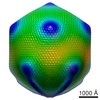
ジャーナル: PLoS Biol / 年: 2009 タイトル: Structural studies of the giant mimivirus. 著者: Chuan Xiao / Yurii G Kuznetsov / Siyang Sun / Susan L Hafenstein / Victor A Kostyuchenko / Paul R Chipman / Marie Suzan-Monti / Didier Raoult / Alexander McPherson / Michael G Rossmann / 要旨: Mimivirus is the largest known virus whose genome and physical size are comparable to some small bacteria, blurring the boundary between a virus and a cell. Structural studies of Mimivirus have been ...Mimivirus is the largest known virus whose genome and physical size are comparable to some small bacteria, blurring the boundary between a virus and a cell. Structural studies of Mimivirus have been difficult because of its size and long surface fibers. Here we report the use of enzymatic digestions to remove the surface fibers of Mimivirus in order to expose the surface of the viral capsid. Cryo-electron microscopy (cryoEM) and atomic force microscopy were able to show that the 20 icosahedral faces of Mimivirus capsids have hexagonal arrays of depressions. Each depression is surrounded by six trimeric capsomers that are similar in structure to those in many other large, icosahedral double-stranded DNA viruses. Whereas in most viruses these capsomers are hexagonally close-packed with the same orientation in each face, in Mimivirus there are vacancies at the systematic depressions with neighboring capsomers differing in orientation by 60 degrees . The previously observed starfish-shaped feature is well-resolved and found to be on each virus particle and is associated with a special pentameric vertex. The arms of the starfish fit into the gaps between the five faces surrounding the unique vertex, acting as a seal. Furthermore, the enveloped nucleocapsid is accurately positioned and oriented within the capsid with a concave surface facing the unique vertex. Thus, the starfish-shaped feature and the organization of the nucleocapsid might regulate the delivery of the genome to the host. The structure of Mimivirus, as well as the various fiber components observed in the virus, suggests that the Mimivirus genome includes genes derived from both eukaryotic and prokaryotic organisms. The three-dimensional cryoEM reconstruction reported here is of a virus with a volume that is one order of magnitude larger than any previously reported molecular assembly studied at a resolution of equal to or better than 65 Angstroms.
pH: 7.4 / 詳細: PBS (137 mM NaCl, 10 mM Phosphate, 2.7 mM KCl)
グリッド
詳細: 200 Copper grid
凍結
凍結剤: ETHANE / チャンバー内温度: 80 K / 装置: REICHERT-JUNG PLUNGER 詳細: Vitrification instrument: Reichert plunger. Using Quantifoil S7 slash 2 grid 手法: Blot for 1 seconds before plunging
-
電子顕微鏡法
顕微鏡
FEI/PHILIPS CM300FEG/T
温度
平均: 80 K
アライメント法
Legacy - 非点収差: objective lens astigmatism was corrected at 98,000 times magnification
撮影
カテゴリ: FILM / フィルム・検出器のモデル: KODAK SO-163 FILM デジタル化 - スキャナー: NIKON SUPER COOLSCAN 9000 デジタル化 - サンプリング間隔: 38.1 µm / 平均電子線量: 20 e/Å2 / 詳細: 6.35 um scanned then bined 6 times to 38.1 um / ビット/ピクセル: 16
 ムービー
ムービー コントローラー


 データを開く
 基本情報
基本情報 マップデータ
 試料
試料 キーワード
キーワード
 Acanthamoeba polyphaga mimivirus (ウイルス)
 データ登録者
 引用
引用
 構造の表示
構造の表示 ムービービューア
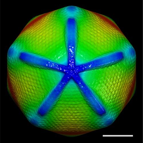
 ダウンロードとリンク
 http://ftp.pdbj.org/pub/emdb/structures/EMD-5039
 Z (Sec.)
 Y (Row.)
 X (Col.)


 試料の構成要素
 Acanthamoeba polyphaga (多食アメーバ) / 別称: PROTOZOA
 解析
解析 電子顕微鏡法
 FIELD EMISSION GUN