 ムービー
ムービー コントローラー


+ データを開く

- 基本情報
基本情報
| 登録情報 | データベース: EMDB / ID: EMD-5033 | |||||||||
|---|---|---|---|---|---|---|---|---|---|---|
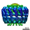
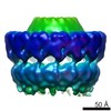
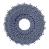
| タイトル | Structure of a type IV secretion system core complex | |||||||||
 マップデータ マップデータ | volume | |||||||||
 試料 試料 |
| |||||||||
 キーワード キーワード | bacterial secretion / type IV secretion / vir / tra | |||||||||
| 手法 | 単粒子再構成法 / ネガティブ染色法 / 解像度: 19.0 Å | |||||||||
 データ登録者 データ登録者 | Fronzes R / Schafer E / Wang L / Saibil H / Orlova E / Waksman G | |||||||||
 引用 引用 | ジャーナル: Science / 年: 2009 タイトル: Structure of a type IV secretion system core complex. 著者: Rémi Fronzes / Eva Schäfer / Luchun Wang / Helen R Saibil / Elena V Orlova / Gabriel Waksman /  要旨: Type IV secretion systems (T4SSs) are important virulence factors used by Gram-negative bacterial pathogens to inject effectors into host cells or to spread plasmids harboring antibiotic resistance ...Type IV secretion systems (T4SSs) are important virulence factors used by Gram-negative bacterial pathogens to inject effectors into host cells or to spread plasmids harboring antibiotic resistance genes. We report the 15 angstrom resolution cryo-electron microscopy structure of the core complex of a T4SS. The core complex is composed of three proteins, each present in 14 copies and forming a approximately 1.1-megadalton two-chambered, double membrane-spanning channel. The structure is double-walled, with each component apparently spanning a large part of the channel. The complex is open on the cytoplasmic side and constricted on the extracellular side. Overall, the T4SS core complex structure is different in both architecture and composition from the other known double membrane-spanning secretion system that has been structurally characterized. | |||||||||
| 履歴 |
|
- 構造の表示
構造の表示
| ムービー |
 ムービービューア ムービービューア |
|---|---|
| 構造ビューア | EMマップ: SurfViewMolmilJmol/JSmol |
| 添付画像 |
 UCSF Chimera
- ダウンロードとリンク
-EMDBアーカイブ
| マップデータ | emd_5033.map.gz | 1.5 MB | EMDBマップデータ形式 | |
|---|---|---|---|---|
| ヘッダ (付随情報) | emd-5033-v30.xmlemd-5033.xml | 11.9 KB 11.9 KB | 表示 表示 | EMDBヘッダ |
| 画像 | 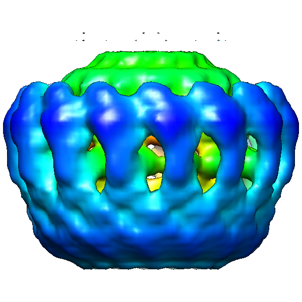 emd_5033_1.png emd_5033_1.png | 203.9 KB | ||
| アーカイブディレクトリ |  http://ftp.pdbj.org/pub/emdb/structures/EMD-5033ftp://ftp.pdbj.org/pub/emdb/structures/EMD-5033 http://ftp.pdbj.org/pub/emdb/structures/EMD-5033ftp://ftp.pdbj.org/pub/emdb/structures/EMD-5033 | HTTPS FTP |
-検証レポート
| 文書・要旨 | emd_5033_validation.pdf.gz | 78.4 KB | 表示 | EMDB検証レポート |
|---|---|---|---|---|
| 文書・詳細版 | emd_5033_full_validation.pdf.gz | 77.5 KB | 表示 | |
| XML形式データ | emd_5033_validation.xml.gz | 493 B | 表示 | |
| アーカイブディレクトリ | https://ftp.pdbj.org/pub/emdb/validation_reports/EMD-5033ftp://ftp.pdbj.org/pub/emdb/validation_reports/EMD-5033 | HTTPS FTP |
-関連構造データ
-リンク
| EMDBのページ | EMDB (EBI/PDBe) / EMDataResource |
|---|
-マップ
| ファイル | ダウンロード / ファイル: emd_5033.map.gz / 形式: CCP4 / 大きさ: 29.8 MB / タイプ: IMAGE STORED AS FLOATING POINT NUMBER (4 BYTES) | ||||||||||||||||||||||||||||||||||||||||||||||||||||||||||||||||||||
|---|---|---|---|---|---|---|---|---|---|---|---|---|---|---|---|---|---|---|---|---|---|---|---|---|---|---|---|---|---|---|---|---|---|---|---|---|---|---|---|---|---|---|---|---|---|---|---|---|---|---|---|---|---|---|---|---|---|---|---|---|---|---|---|---|---|---|---|---|---|
| 注釈 | volume | ||||||||||||||||||||||||||||||||||||||||||||||||||||||||||||||||||||
| 投影像・断面図 | 画像のコントロール
画像は Spider により作成 | ||||||||||||||||||||||||||||||||||||||||||||||||||||||||||||||||||||
| ボクセルのサイズ | X=Y=Z: 2.5 Å | ||||||||||||||||||||||||||||||||||||||||||||||||||||||||||||||||||||
| 密度 |
| ||||||||||||||||||||||||||||||||||||||||||||||||||||||||||||||||||||
| 対称性 | 空間群: 1 | ||||||||||||||||||||||||||||||||||||||||||||||||||||||||||||||||||||
| 詳細 | EMDB XML:
CCP4マップ ヘッダ情報:
| ||||||||||||||||||||||||||||||||||||||||||||||||||||||||||||||||||||
 Z (Sec.)
 Y (Row.)
 X (Col.)


-添付データ
- 試料の構成要素
-全体 : traN/traO/traF complex encoded by pKM101 Digested with 0.002 mg m...
| 全体 | 名称: traN/traO/traF complex encoded by pKM101 Digested with 0.002 mg ml-1 of trypsin for 30 min at 4 degrees Celsius. |
|---|---|
| 要素 |
|
-超分子 #1000: traN/traO/traF complex encoded by pKM101 Digested with 0.002 mg m...
| 超分子 | 名称: traN/traO/traF complex encoded by pKM101 Digested with 0.002 mg ml-1 of trypsin for 30 min at 4 degrees Celsius. タイプ: sample / ID: 1000 / 詳細: monodisperse / 集合状態: 14-mer / Number unique components: 3 |
|---|---|
| 分子量 | 実験値: 868 KDa / 理論値: 700 KDa / 手法: gel filtration |
-分子 #1: traF
| 分子 | 名称: traF / タイプ: protein_or_peptide / ID: 1 / Name.synonym: traF / コピー数: 14 / 集合状態: 14-mer / 組換発現: Yes |
|---|---|
| 由来(天然) | 株: BL21 / 細胞: Escherichia coli / 細胞中の位置: inner membrane |
| 分子量 | 理論値: 40 KDa |
| 組換発現 | 生物種:  |
-分子 #2: traO
| 分子 | 名称: traO / タイプ: protein_or_peptide / ID: 2 / Name.synonym: traO / コピー数: 14 / 集合状態: 14-mer / 組換発現: Yes |
|---|---|
| 由来(天然) | 株: BL21 / 細胞: Escherichia coli / 細胞中の位置: outer membrane |
| 分子量 | 理論値: 30 KDa |
| 組換発現 | 生物種: |
-分子 #3: traN
| 分子 | 名称: traN / タイプ: protein_or_peptide / ID: 3 / Name.synonym: traN / コピー数: 14 / 集合状態: 14-mer / 組換発現: Yes |
|---|---|
| 由来(天然) | 株: BL21 / 細胞: Escherichia coli / 細胞中の位置: outer membrane |
| 分子量 | 理論値: 5 KDa |
| 組換発現 | 生物種: |
-実験情報
-構造解析
| 手法 | ネガティブ染色法 |
|---|---|
 解析 解析 | 単粒子再構成法 |
| 試料の集合状態 | particle |
-試料調製
| 濃度 | 0.5 mg/mL |
|---|---|
| 緩衝液 | 詳細: 50 mM Tris-HCL, 200 mM NaCl, 10 mM LDAO |
| 染色 | タイプ: NEGATIVE / 詳細: 2% uranyl acetate |
| グリッド | 詳細: carbon coated copper grids |
| 凍結 | 凍結剤: NONE / 装置: OTHER |
- 電子顕微鏡法
| 顕微鏡 | FEI TECNAI 12 |
|---|---|
| 温度 | 最低: 293 K / 最高: 293 K / 平均: 293 K |
| 日付 | 2008年1月1日 |
| 撮影 | カテゴリ: FILM / フィルム・検出器のモデル: KODAK SO-163 FILM / デジタル化 - スキャナー: ZEISS SCAI / デジタル化 - サンプリング間隔: 7 µm / 実像数: 22 / 平均電子線量: 20 e/Å2 / Od range: 2 / ビット/ピクセル: 8 |
| 電子線 | 加速電圧: 120 kV / 電子線源: TUNGSTEN HAIRPIN |
| 電子光学系 | 倍率(補正後): 42000 / 照射モード: FLOOD BEAM / 撮影モード: BRIGHT FIELD / Cs: 2.2 mm / 最大 デフォーカス(公称値): 2.0 µm / 最小 デフォーカス(公称値): 0.8 µm / 倍率(公称値): 42000 |
| 試料ステージ | 試料ホルダー: side entry room temperature / 試料ホルダーモデル: OTHER |
-画像解析
| 最終 再構成 | アルゴリズム: OTHER / 解像度のタイプ: BY AUTHOR / 解像度: 19.0 Å / 解像度の算出法: FSC 0.5 CUT-OFF / ソフトウェア - 名称: imagic / 詳細: final maps were calculated from 2201 particles / 使用した粒子像数: 2201 |
|---|---|
| 最終 2次元分類 | クラス数: 150 |
