National Institutes of Health/National Institute of General Medical Sciences (NIH/NIGMS)
GM058067
United States
H2020 Marie Curie Actions of the European Commission
893196
European Union
Citation
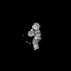
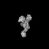
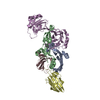
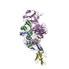
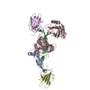
Journal: Nat Commun / Year: 2025 Title: Structures of TGF-β with betaglycan and signaling receptors reveal mechanisms of complex assembly and signaling. Authors: Łukasz Wieteska / Alexander B Taylor / Emma Punch / Jonathan A Coleman / Isabella O Conway / Yeu-Farn Lin / Chang-Hyeock Byeon / Cynthia S Hinck / Troy Krzysiak / Rieko Ishima / Fernando ...Authors: Łukasz Wieteska / Alexander B Taylor / Emma Punch / Jonathan A Coleman / Isabella O Conway / Yeu-Farn Lin / Chang-Hyeock Byeon / Cynthia S Hinck / Troy Krzysiak / Rieko Ishima / Fernando López-Casillas / Peter Cherepanov / Daniel J Bernard / Caroline S Hill / Andrew P Hinck / Abstract: Betaglycan (BG) is a transmembrane co-receptor of the transforming growth factor-β (TGF-β) family of signaling ligands. It is essential for embryonic development, tissue homeostasis and fertility ...Betaglycan (BG) is a transmembrane co-receptor of the transforming growth factor-β (TGF-β) family of signaling ligands. It is essential for embryonic development, tissue homeostasis and fertility in adults. It functions by enabling binding of the three TGF-β isoforms to their signaling receptors and is additionally required for inhibin A (InhA) activity. Despite its requirement for the functions of TGF-βs and InhA in vivo, structural information explaining BG ligand selectivity and its mechanism of action is lacking. Here, we determine the structure of TGF-β bound both to BG and the signaling receptors, TGFBR1 and TGFBR2. We identify key regions responsible for ligand engagement, which has revealed binding interfaces that differ from those described for the closely related co-receptor of the TGF-β family, endoglin, thus demonstrating remarkable evolutionary adaptation to enable ligand selectivity. Finally, we provide a structural explanation for the hand-off mechanism underlying TGF-β signal potentiation.
In the structure databanks used in Yorodumi, some data are registered as the other names, "COVID-19 virus" and "2019-nCoV". Here are the details of the virus and the list of structure data.
Jan 31, 2019. EMDB accession codes are about to change! (news from PDBe EMDB page)
EMDB accession codes are about to change! (news from PDBe EMDB page)
The allocation of 4 digits for EMDB accession codes will soon come to an end. Whilst these codes will remain in use, new EMDB accession codes will include an additional digit and will expand incrementally as the available range of codes is exhausted. The current 4-digit format prefixed with “EMD-” (i.e. EMD-XXXX) will advance to a 5-digit format (i.e. EMD-XXXXX), and so on. It is currently estimated that the 4-digit codes will be depleted around Spring 2019, at which point the 5-digit format will come into force.
The EM Navigator/Yorodumi systems omit the EMD- prefix.
Related info.:Q: What is EMD? / ID/Accession-code notation in Yorodumi/EM Navigator
Yorodumi is a browser for structure data from EMDB, PDB, SASBDB, etc.
This page is also the successor to EM Navigator detail page, and also detail information page/front-end page for Omokage search.
The word "yorodu" (or yorozu) is an old Japanese word meaning "ten thousand". "mi" (miru) is to see.
Related info.:EMDB / PDB / SASBDB / Comparison of 3 databanks / Yorodumi Search / Aug 31, 2016. New EM Navigator & Yorodumi / Yorodumi Papers / Jmol/JSmol / Function and homology information / Changes in new EM Navigator and Yorodumi
 Movie
Movie Controller


 Open data
 Basic information
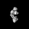
 Map data
 Sample
 Keywords
 Function and homology information
 Homo sapiens (human)
 Authors
 United States, European Union, 2 items
  Citation


 Structure visualization
 Downloads & links
 emd_50326.png
 http://ftp.pdbj.org/pub/emdb/structures/EMD-50326
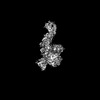


 Z (Sec.)
 Y (Row.)
 X (Col.)


 Sample components
 Processing
 Electron microscopy
 FIELD EMISSION GUN

