 Movie
Movie Controller

[English] 日本語
 Yorodumi
- EMDB-5026: Cryo-negative stain structure of the yeast transcription factor T... -
+ Open data

- Basic information
| Entry | Database: EMDB / ID: EMD-5026 | |||||||||
|---|---|---|---|---|---|---|---|---|---|---|
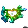
| Title | Cryo-negative stain structure of the yeast transcription factor TFIID at 20 A resolution | |||||||||
 Map data Map data | Volume of yeast TFIID filtered to 20 A resolution | |||||||||
 Sample Sample |
| |||||||||
 Keywords Keywords | Transcription factor TFIID / cryo electron microscopy / yeast | |||||||||
| Method | single particle reconstruction / cryo EM / negative staining / Resolution: 22.0 Å | |||||||||
 Authors Authors | Papai G / Tripathi MK / Ruhlmann C / Crucifix C / Jennings JL / Link AJ / Weil PA / Schultz P | |||||||||
 Citation Citation | Journal: Structure / Year: 2009 Title: Mapping the initiator binding Taf2 subunit in the structure of hydrated yeast TFIID. Authors: Gabor Papai / Manish K Tripathi / Christine Ruhlmann / Sebastiaan Werten / Corinne Crucifix / P Anthony Weil / Patrick Schultz /  Abstract: The general transcription factor TFIID is a large multisubunit complex required for the transcription of most protein-encoding genes by RNA polymerase II. Taking advantage of a TFIID preparation ...The general transcription factor TFIID is a large multisubunit complex required for the transcription of most protein-encoding genes by RNA polymerase II. Taking advantage of a TFIID preparation partially depleted in the initiator-binding Taf2p subunit, we determined the conformational and biochemical variations of the complex by electron tomography and cryo-electron microscopy of single molecules. Image analysis revealed the extent of conformational flexibility of the complex and the selection of the most homogeneous TFIID subpopulation allowed us to determine an improved structural model at 23 Angstroms resolution. This study also identified two subpopulations of Taf2p-containing and Taf2p-depleted TFIID molecules. By comparing these two TFIID species we could infer the position of Taf2p, which was confirmed by immunolabeling using a subunit-specific antibody. Mapping the position of this crucial subunit in the vicinity of Taf1p and of TBP sheds new light on its role in promoter recognition. | |||||||||
| History |
|
- Structure visualization
| Movie |
 Movie viewer Movie viewer |
|---|---|
| Structure viewer | EM map: SurfViewMolmilJmol/JSmol |
| Supplemental images |

- Downloads & links
-EMDB archive
| Map data | emd_5026.map.gz | 14.2 MB | EMDB map data format | |
|---|---|---|---|---|
| Header (meta data) | emd-5026-v30.xmlemd-5026.xml | 10.2 KB 10.2 KB | Display Display | EMDB header |
| Images | 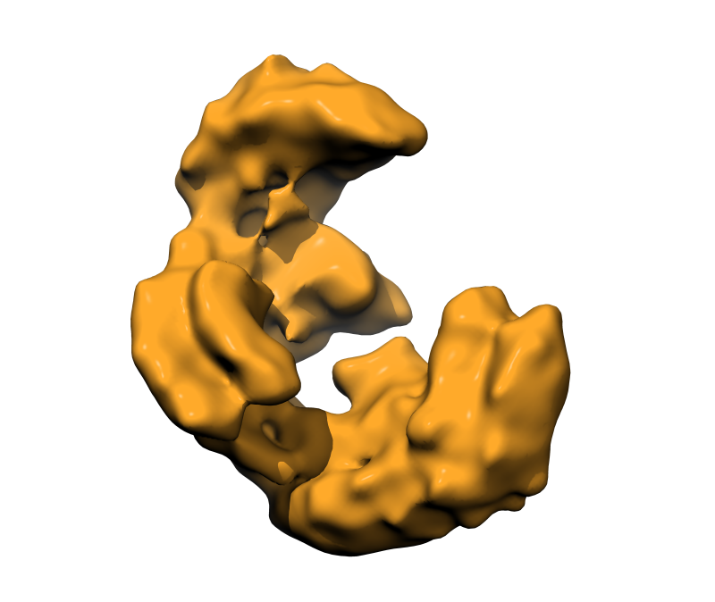 emd_5026_1.png emd_5026_1.png | 183.2 KB | ||
| Archive directory |  http://ftp.pdbj.org/pub/emdb/structures/EMD-5026ftp://ftp.pdbj.org/pub/emdb/structures/EMD-5026 http://ftp.pdbj.org/pub/emdb/structures/EMD-5026ftp://ftp.pdbj.org/pub/emdb/structures/EMD-5026 | HTTPS FTP |
-Related structure data
| Similar structure data |
|---|


-Links
| EMDB pages | EMDB (EBI/PDBe) / EMDataResource |
|---|
-Map
| File | Download / File: emd_5026.map.gz / Format: CCP4 / Size: 15.3 MB / Type: IMAGE STORED AS FLOATING POINT NUMBER (4 BYTES) | ||||||||||||||||||||||||||||||||||||||||||||||||||||||||||||||||||||
|---|---|---|---|---|---|---|---|---|---|---|---|---|---|---|---|---|---|---|---|---|---|---|---|---|---|---|---|---|---|---|---|---|---|---|---|---|---|---|---|---|---|---|---|---|---|---|---|---|---|---|---|---|---|---|---|---|---|---|---|---|---|---|---|---|---|---|---|---|---|
| Annotation | Volume of yeast TFIID filtered to 20 A resolution | ||||||||||||||||||||||||||||||||||||||||||||||||||||||||||||||||||||
| Projections & slices | Image control
Images are generated by Spider. | ||||||||||||||||||||||||||||||||||||||||||||||||||||||||||||||||||||
| Voxel size | X=Y=Z: 2.54 Å | ||||||||||||||||||||||||||||||||||||||||||||||||||||||||||||||||||||
| Density |
| ||||||||||||||||||||||||||||||||||||||||||||||||||||||||||||||||||||
| Symmetry | Space group: 1 | ||||||||||||||||||||||||||||||||||||||||||||||||||||||||||||||||||||
| Details | EMDB XML:
CCP4 map header:
| ||||||||||||||||||||||||||||||||||||||||||||||||||||||||||||||||||||
 Z (Sec.)
 Y (Row.)
 X (Col.)


-Supplemental data
- Sample components
-Entire : TAp-tag purified yeast TFIID
| Entire | Name: TAp-tag purified yeast TFIID |
|---|---|
| Components |
|
-Supramolecule #1000: TAp-tag purified yeast TFIID
| Supramolecule | Name: TAp-tag purified yeast TFIID / type: sample / ID: 1000 / Oligomeric state: monomeric / Number unique components: 1 |
|---|---|
| Molecular weight | Experimental: 900 KDa / Theoretical: 900 KDa |
-Macromolecule #1: Transcription factor TFIID
| Macromolecule | Name: Transcription factor TFIID / type: protein_or_peptide / ID: 1 / Name.synonym: TFIID / Number of copies: 1 / Oligomeric state: monomer / Recombinant expression: Yes / Database: NCBI |
|---|---|
| Source (natural) | Strain: YLSTAF1 / Cell: yeast / Location in cell: nuclear protein |
| Molecular weight | Experimental: 900 MDa / Theoretical: 900 MDa |
-Experimental details
-Structure determination
| Method | negative staining, cryo EM |
|---|---|
 Processing Processing | single particle reconstruction |
| Aggregation state | particle |
-Sample preparation
| Concentration | 1.4 mg/mL |
|---|---|
| Buffer | pH: 8 / Details: 10 mM Tris-HCL, 300 mM NaCl, 20% glycerol |
| Staining | Type: NEGATIVE Details: Thin carbon film with adsorbed protein were floated on 2% w/v uranyl acetate for 30 seconds. |
| Grid | Details: 400 mesh copper/rhodium grid with holey carbon |
| Vitrification | Cryogen name: ETHANE / Instrument: HOMEMADE PLUNGER / Details: Vitrification instrument: house made / Method: blot for about 2 seconds before plunging |
- Electron microscopy
| Microscope | FEI TECNAI F20 |
|---|---|
| Temperature | Average: 90 K |
| Date | Sep 19, 2006 |
| Image recording | Category: FILM / Film or detector model: KODAK SO-163 FILM / Digitization - Scanner: PRIMESCAN / Digitization - Sampling interval: 5.1 µm / Number real images: 121 / Average electron dose: 15 e/Å2 / Od range: 1.4 / Bits/pixel: 16 |
| Electron beam | Acceleration voltage: 200 kV / Electron source:  FIELD EMISSION GUN FIELD EMISSION GUN |
| Electron optics | Calibrated magnification: 40080 / Illumination mode: FLOOD BEAM / Imaging mode: BRIGHT FIELD / Cs: 2.0 mm / Nominal defocus max: 1.96 µm / Nominal defocus min: 0.137 µm / Nominal magnification: 40000 |
| Sample stage | Specimen holder: Side entry / Specimen holder model: GATAN LIQUID NITROGEN |
| Experimental equipment |  Model: Tecnai F20 / Image courtesy: FEI Company |
-Image processing
| Details | Most homogeneous particles were sorted out by using several starting models obtained by electron tomography. |
|---|---|
| CTF correction | Details: phase flipping on each particle from one micrograph |
| Final reconstruction | Algorithm: OTHER / Resolution.type: BY AUTHOR / Resolution: 22.0 Å / Resolution method: FSC 0.5 CUT-OFF / Software - Name: IMAGIC, SPIDER / Number images used: 10205 |
| Final two d classification | Number classes: 700 |
