 Movie
Movie Controller

[English] 日本語
 Yorodumi
- EMDB-50134: Electron tomogram of ER-ER/nuclear envelope junction of HeLa cell... -
+ Open data

- Basic information
| Entry | 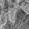 | |||||||||
|---|---|---|---|---|---|---|---|---|---|---|
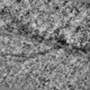
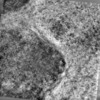
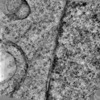
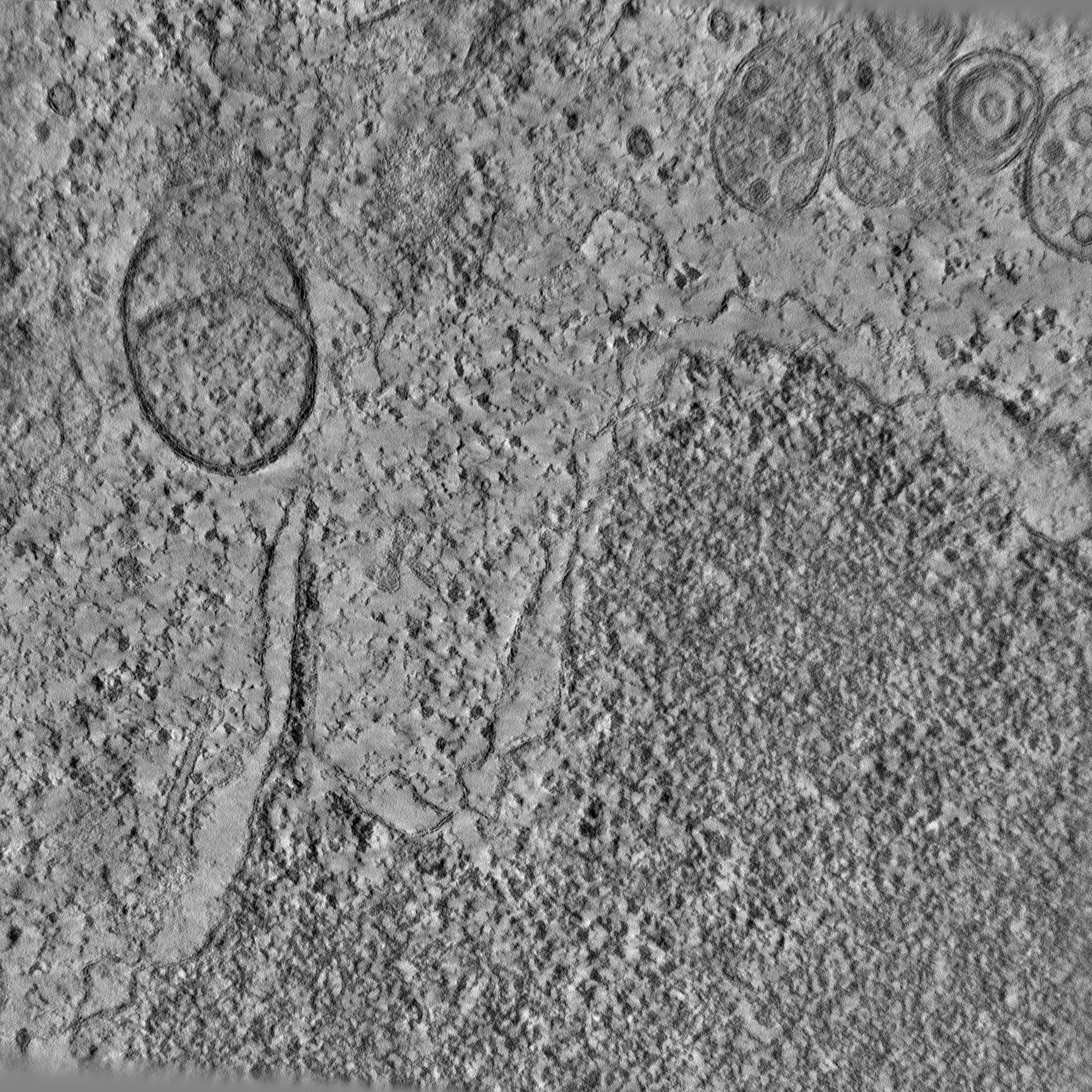
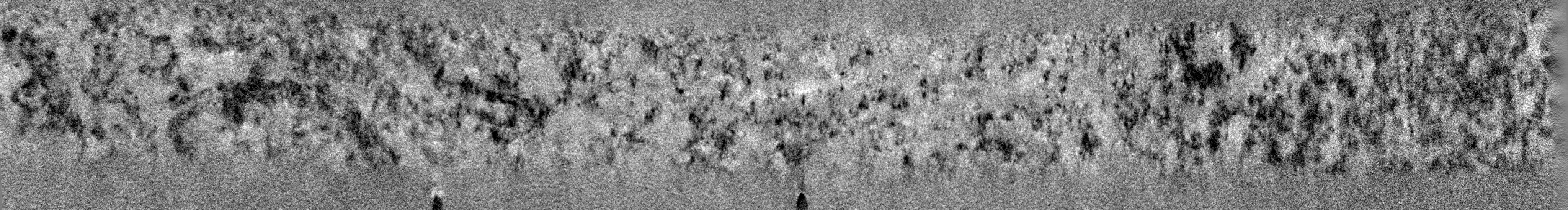
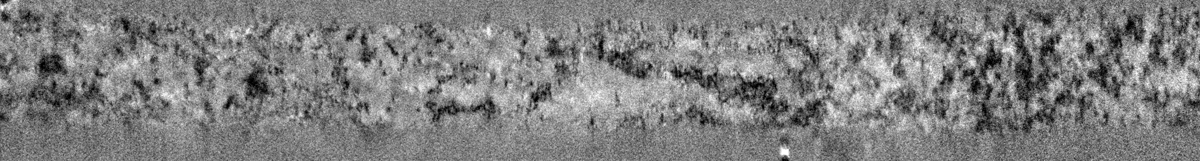
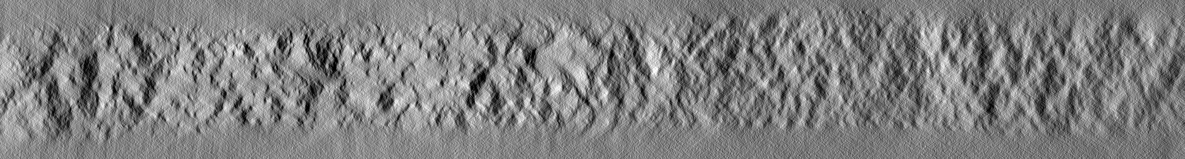
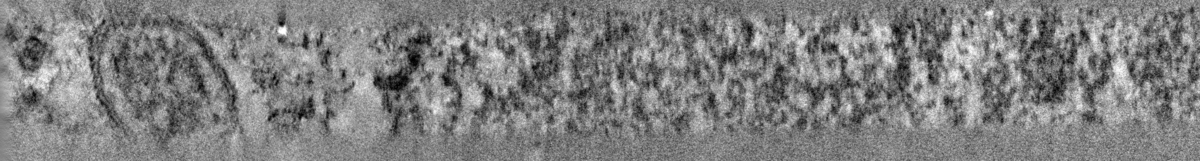
| Title | Electron tomogram of ER-ER/nuclear envelope junction of HeLa cell in late anaphase | |||||||||
 Map data Map data | ||||||||||
 Sample Sample |
| |||||||||
 Keywords Keywords | Endoplasmic reticulum / Nuclear envelope / Organelle contact site / Membrane junction / CELL CYCLE | |||||||||
| Biological species |  Homo sapiens (human) Homo sapiens (human) | |||||||||
| Method | electron tomography / cryo EM / negative staining | |||||||||
 Authors Authors | Bragulat-Teixidor H / Otsuka S | |||||||||
| Funding support |  Austria, 2 items Austria, 2 items
| |||||||||
 Citation Citation | Journal: EMBO Rep / Year: 2024 Title: The endoplasmic reticulum connects to the nucleus by constricted junctions that mature after mitosis. Authors: Helena Bragulat-Teixidor / Keisuke Ishihara / Gréta Martina Szücs / Shotaro Otsuka /  Abstract: Junctions between the endoplasmic reticulum (ER) and the outer membrane of the nuclear envelope (NE) physically connect both organelles. These ER-NE junctions are essential for supplying the NE with ...Junctions between the endoplasmic reticulum (ER) and the outer membrane of the nuclear envelope (NE) physically connect both organelles. These ER-NE junctions are essential for supplying the NE with lipids and proteins synthesized in the ER. However, little is known about the structure of these ER-NE junctions. Here, we systematically study the ultrastructure of ER-NE junctions in cryo-fixed mammalian cells staged in anaphase, telophase, and interphase by correlating live cell imaging with three-dimensional electron microscopy. Our results show that ER-NE junctions in interphase cells have a pronounced hourglass shape with a constricted neck of 7-20 nm width. This morphology is significantly distinct from that of junctions within the ER network, and their morphology emerges as early as telophase. The highly constricted ER-NE junctions are seen in several mammalian cell types, but not in budding yeast. We speculate that the unique and highly constricted ER-NE junctions are regulated via novel mechanisms that contribute to ER-to-NE lipid and protein traffic in higher eukaryotes. | |||||||||
| History |
|
- Structure visualization
| Supplemental images |
|---|

- Downloads & links
-EMDB archive
| Map data | emd_50134.map.gz | 12.3 GB |  EMDB map data format EMDB map data format | |
|---|---|---|---|---|
| Header (meta data) | emd-50134-v30.xmlemd-50134.xml | 9 KB 9 KB | Display Display | EMDB header |
| Images | 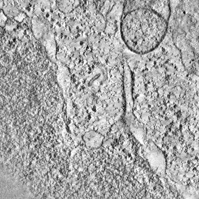 emd_50134.png emd_50134.png | 248.5 KB | ||
| Filedesc metadata | emd-50134.cif.gz | 3.9 KB | ||
| Archive directory |  http://ftp.pdbj.org/pub/emdb/structures/EMD-50134ftp://ftp.pdbj.org/pub/emdb/structures/EMD-50134 http://ftp.pdbj.org/pub/emdb/structures/EMD-50134ftp://ftp.pdbj.org/pub/emdb/structures/EMD-50134 | HTTPS FTP |
-Validation report
| Summary document | emd_50134_validation.pdf.gz | 494.9 KB | Display | EMDB validaton report |
|---|---|---|---|---|
| Full document | emd_50134_full_validation.pdf.gz | 494.4 KB | Display | |
| Data in XML | emd_50134_validation.xml.gz | 4 KB | Display | |
| Data in CIF | emd_50134_validation.cif.gz | 4.6 KB | Display | |
| Arichive directory | https://ftp.pdbj.org/pub/emdb/validation_reports/EMD-50134ftp://ftp.pdbj.org/pub/emdb/validation_reports/EMD-50134 | HTTPS FTP |
-Related structure data
-Links
| EMDB pages | EMDB (EBI/PDBe) / EMDataResource |
|---|
-Map
| File | Download / File: emd_50134.map.gz / Format: CCP4 / Size: 17.2 GB / Type: IMAGE STORED AS SIGNED INTEGER (2 BYTES) | ||||||||||||||||||||||||||||||||
|---|---|---|---|---|---|---|---|---|---|---|---|---|---|---|---|---|---|---|---|---|---|---|---|---|---|---|---|---|---|---|---|---|---|
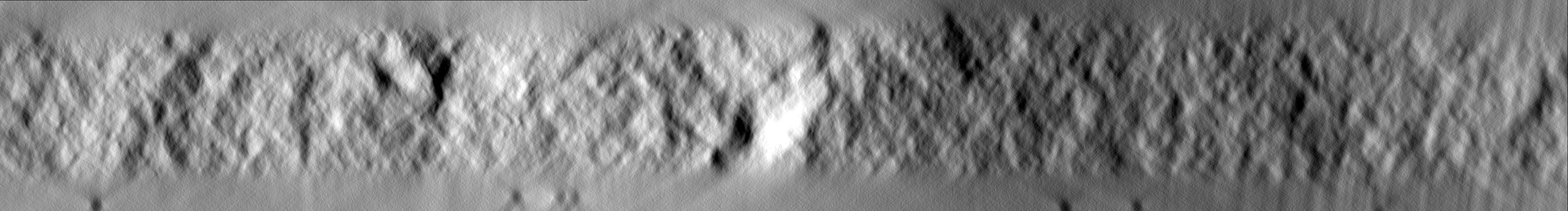
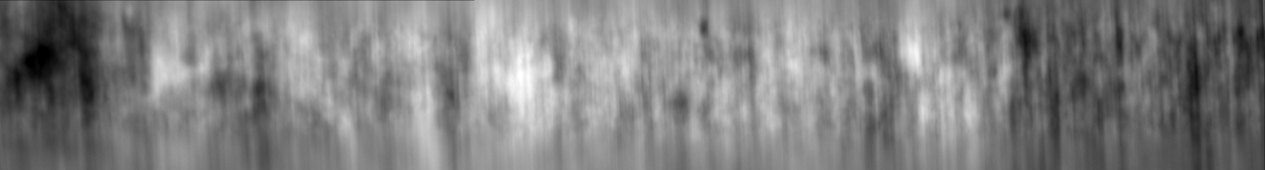
| Projections & slices | Image control
Images are generated by Spider. generated in cubic-lattice coordinate | ||||||||||||||||||||||||||||||||
| Voxel size | X=Y=Z: 4.51 Å | ||||||||||||||||||||||||||||||||
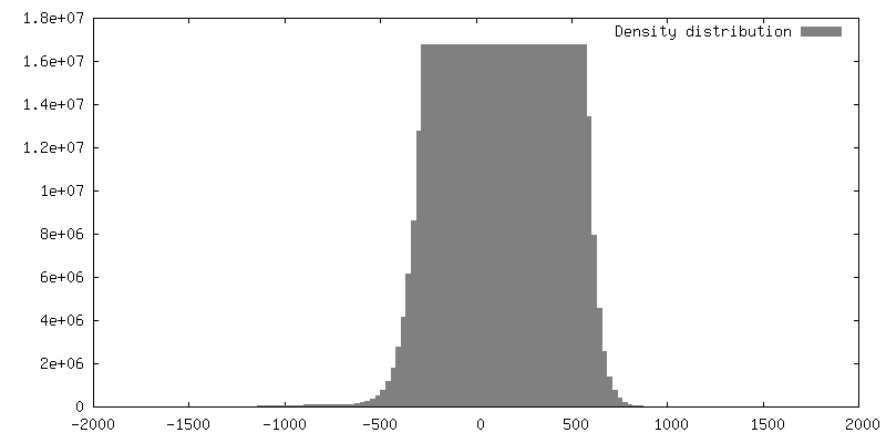
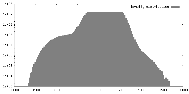
| Density |
| ||||||||||||||||||||||||||||||||
| Symmetry | Space group: 1 | ||||||||||||||||||||||||||||||||
| Details | EMDB XML:
|
 Z (Sec.)
 Y (Row.)
 X (Col.)


-Supplemental data
- Sample components
-Entire : HeLa Kyoto cell
| Entire | Name: HeLa Kyoto cell |
|---|---|
| Components |
|
-Supramolecule #1: HeLa Kyoto cell
| Supramolecule | Name: HeLa Kyoto cell / type: cell / ID: 1 / Parent: 0 |
|---|---|
| Source (natural) | Organism: Homo sapiens (human) / Strain: HeLa |
-Experimental details
-Structure determination
| Method | negative staining, cryo EM |
|---|---|
 Processing Processing | electron tomography |
| Aggregation state | cell |
-Sample preparation
| Buffer | pH: 7.4 Details: DMEM without Riboflavin and Phenol Red, containing 10% FBS, 1% Pen/Strep, and 50 nM SiR-DNA |
|---|---|
| Staining | Type: NEGATIVE / Material: Uranyl acetate and lead citrate |
| Sugar embedding | Material: Agar 100 Epoxy resin Details: Frozen cells were substituted in 0.1% uranyl acetate (UA), 2% Osmium tetroxide and 5% H2O in acetone following this temperature ramp: -90 C to -80 C for 10 hours, -80 C to -30 C for 10 ...Details: Frozen cells were substituted in 0.1% uranyl acetate (UA), 2% Osmium tetroxide and 5% H2O in acetone following this temperature ramp: -90 C to -80 C for 10 hours, -80 C to -30 C for 10 hours, -30 C for 4 hours, -30 C to 0 C for 6 hours, 0 C to 20 C for 4 hours, 20 C for 5-6 hours. |
| Vitrification | Cryogen name: NITROGEN |
| High pressure freezing | Instrument: OTHER Details: High pressure freezing chamber was 1 mm thick, 6.0 mm diameter, with central cavities 5.0 mm x 5.0 mm x 25 um deep. The chamber had been in contact with 1-hexadecene.. The value given for _ ...Details: High pressure freezing chamber was 1 mm thick, 6.0 mm diameter, with central cavities 5.0 mm x 5.0 mm x 25 um deep. The chamber had been in contact with 1-hexadecene.. The value given for _em_high_pressure_freezing.instrument is Leica EM ICE. This is not in a list of allowed values {'OTHER', 'BAL-TEC HPM 010', 'LEICA EM HPM100', 'EMS-002 RAPID IMMERSION FREEZER', 'LEICA EM PACT2', 'LEICA EM PACT'} so OTHER is written into the XML file. |
| Cryo protectant | 20% Ficoll-PM400 |
| Sectioning | Ultramicrotomy - Instrument: Leica Ultracut UCT / Ultramicrotomy - Temperature: 298 K / Ultramicrotomy - Final thickness: 250 |
| Fiducial marker | Manufacturer: Cytodiagnostics / Diameter: 15 nm |
- Electron microscopy
| Microscope | FEI TECNAI F20 |
|---|---|
| Image recording | Film or detector model: FEI EAGLE (4k x 4k) / Average electron dose: 50.0 e/Å2 |
| Electron beam | Acceleration voltage: 200 kV / Electron source:  FIELD EMISSION GUN FIELD EMISSION GUN |
| Electron optics | Illumination mode: FLOOD BEAM / Imaging mode: BRIGHT FIELD / Nominal defocus max: 0.5 µm / Nominal defocus min: 0.2 µm |
| Experimental equipment |  Model: Tecnai F20 / Image courtesy: FEI Company |
-Image processing
| Final reconstruction | Algorithm: BACK PROJECTION / Number images used: 110 |
|---|
