 ムービー
ムービー コントローラー


+ データを開く

- 基本情報
基本情報
| 登録情報 |  | |||||||||
|---|---|---|---|---|---|---|---|---|---|---|
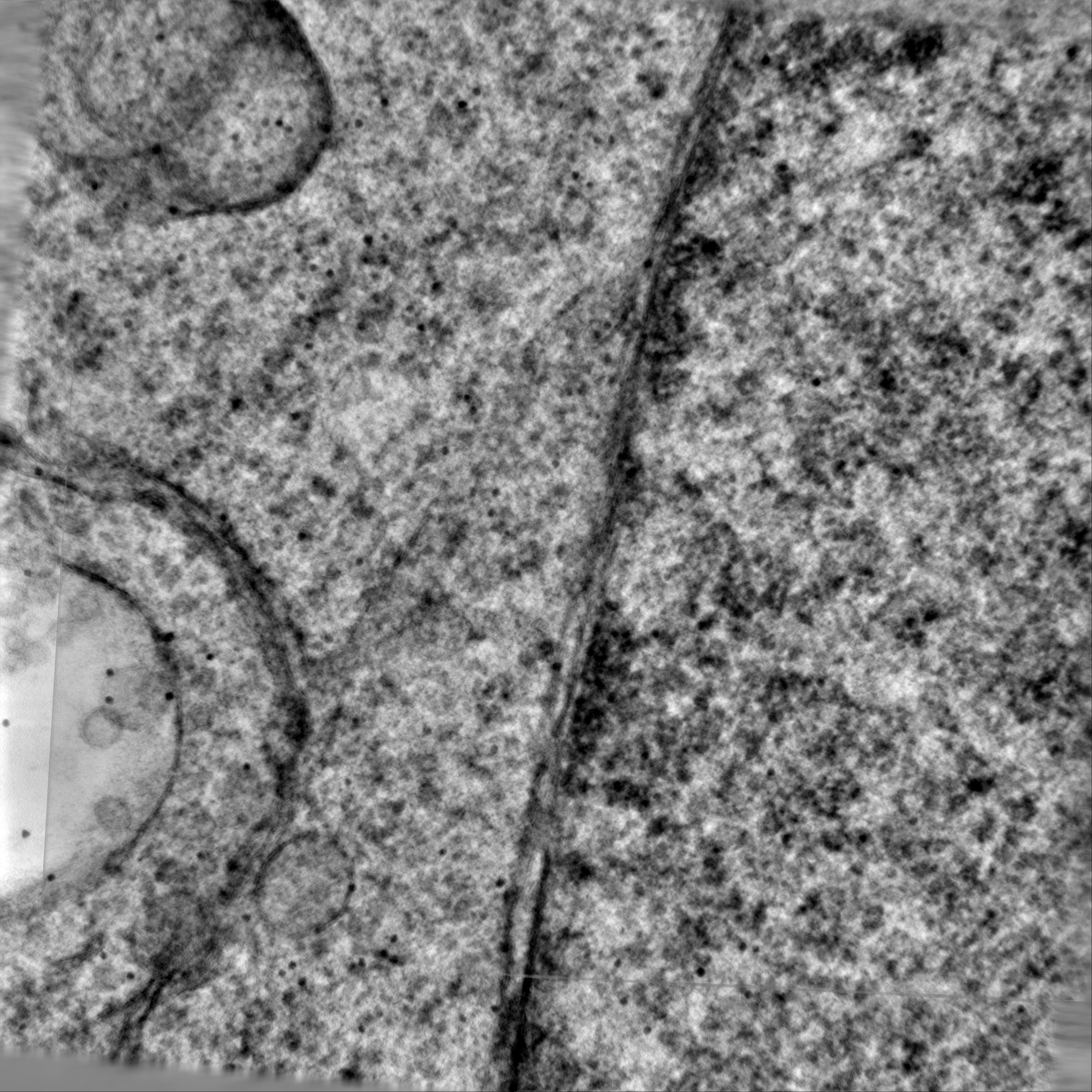
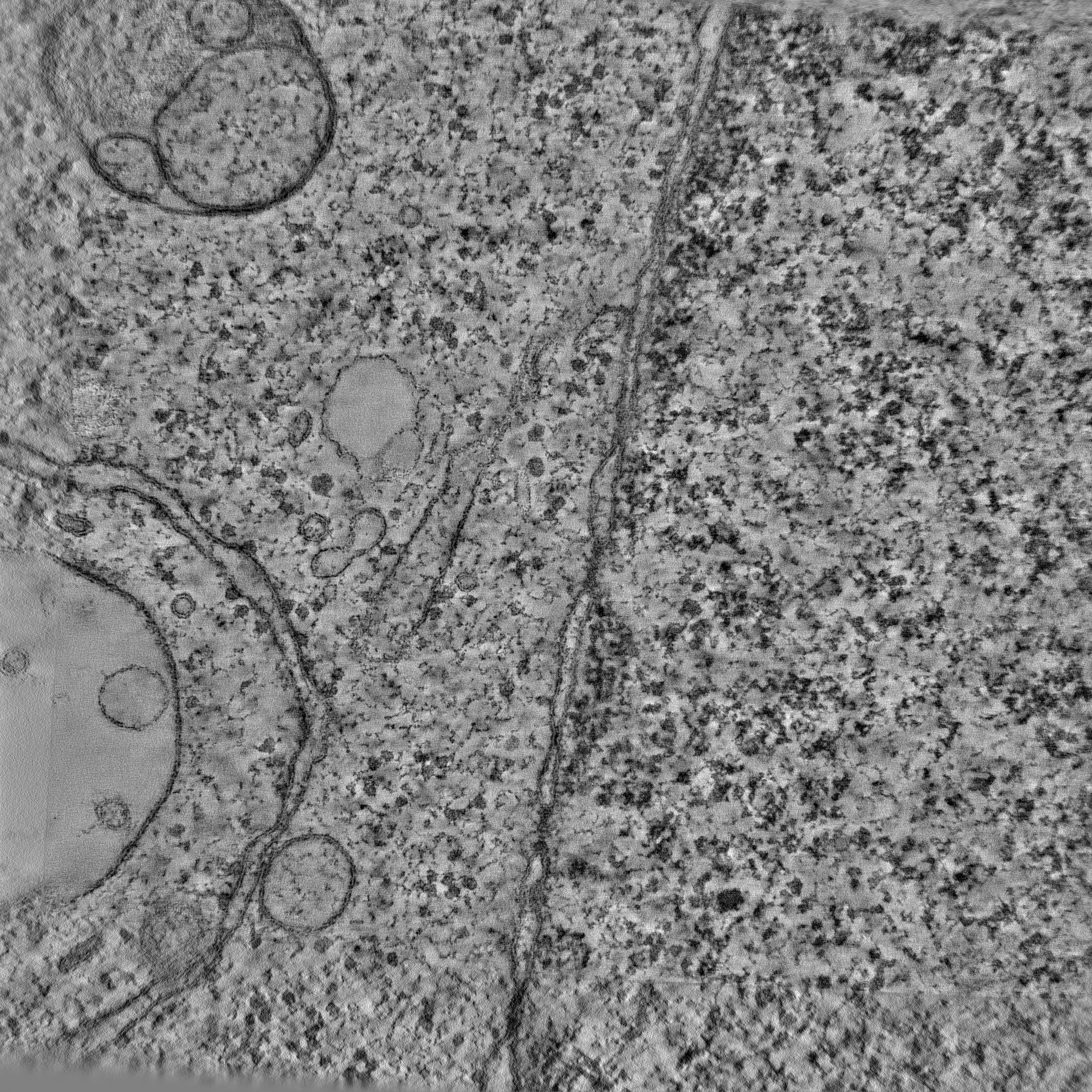
| タイトル | Electron tomogram of ER-ER junction of HeLa cell in interphase | |||||||||
 マップデータ マップデータ | ||||||||||
 試料 試料 |
| |||||||||
 キーワード キーワード | Endoplasmic reticulum / Nuclear envelope / Organelle contact site / Membrane junction / CELL CYCLE | |||||||||
| 生物種 |  Homo sapiens (ヒト) Homo sapiens (ヒト) | |||||||||
| 手法 | 電子線トモグラフィー法 / クライオ電子顕微鏡法 / ネガティブ染色法 | |||||||||
 データ登録者 データ登録者 | Bragulat-Teixidor H / Otsuka S | |||||||||
| 資金援助 |  オーストリア, 2件 オーストリア, 2件
| |||||||||
 引用 引用 | ジャーナル: EMBO Rep / 年: 2024 タイトル: The endoplasmic reticulum connects to the nucleus by constricted junctions that mature after mitosis. 著者: Helena Bragulat-Teixidor / Keisuke Ishihara / Gréta Martina Szücs / Shotaro Otsuka /  要旨: Junctions between the endoplasmic reticulum (ER) and the outer membrane of the nuclear envelope (NE) physically connect both organelles. These ER-NE junctions are essential for supplying the NE with ...Junctions between the endoplasmic reticulum (ER) and the outer membrane of the nuclear envelope (NE) physically connect both organelles. These ER-NE junctions are essential for supplying the NE with lipids and proteins synthesized in the ER. However, little is known about the structure of these ER-NE junctions. Here, we systematically study the ultrastructure of ER-NE junctions in cryo-fixed mammalian cells staged in anaphase, telophase, and interphase by correlating live cell imaging with three-dimensional electron microscopy. Our results show that ER-NE junctions in interphase cells have a pronounced hourglass shape with a constricted neck of 7-20 nm width. This morphology is significantly distinct from that of junctions within the ER network, and their morphology emerges as early as telophase. The highly constricted ER-NE junctions are seen in several mammalian cell types, but not in budding yeast. We speculate that the unique and highly constricted ER-NE junctions are regulated via novel mechanisms that contribute to ER-to-NE lipid and protein traffic in higher eukaryotes. | |||||||||
| 履歴 |
|
- 構造の表示
構造の表示
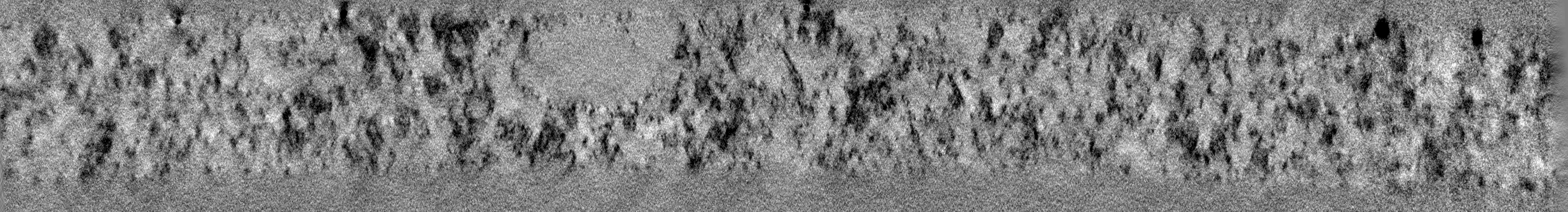
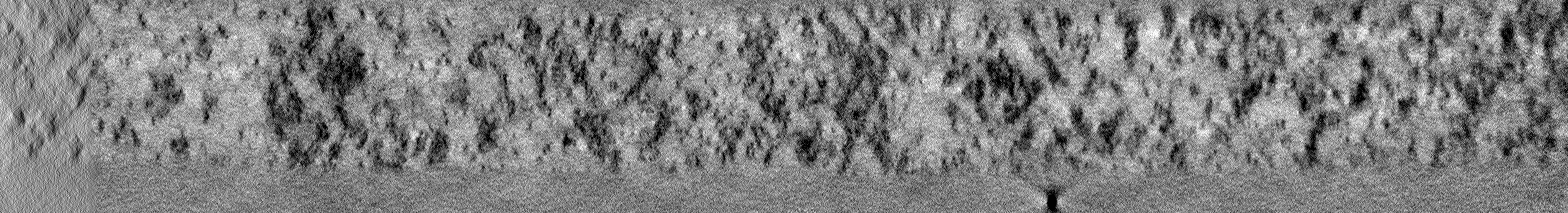
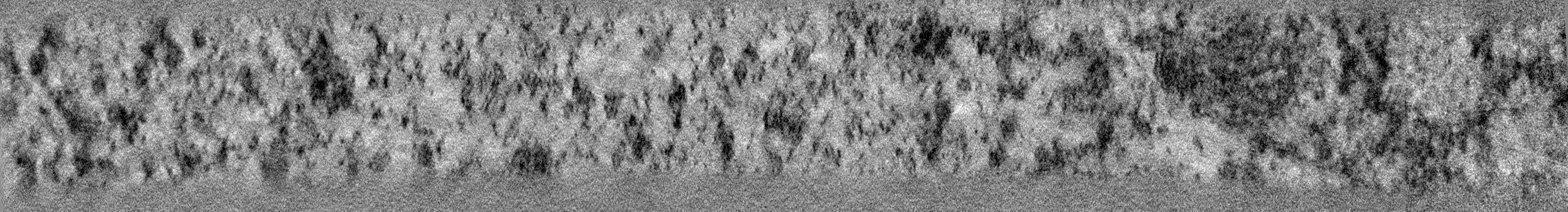
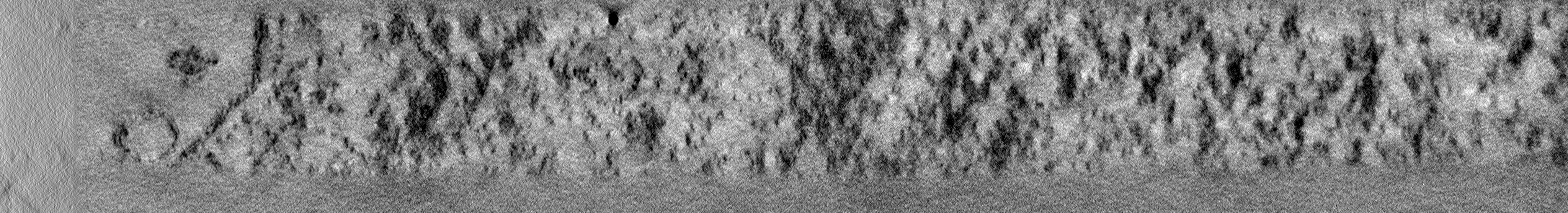
| 添付画像 |
|---|

- ダウンロードとリンク
-EMDBアーカイブ
| マップデータ | emd_50115.map.gz | 12.4 GB |  EMDBマップデータ形式 EMDBマップデータ形式 | |
|---|---|---|---|---|
| ヘッダ (付随情報) | emd-50115-v30.xmlemd-50115.xml | 9 KB 9 KB | 表示 表示 | EMDBヘッダ |
| 画像 | 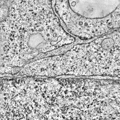 emd_50115.png emd_50115.png | 246.3 KB | ||
| Filedesc metadata | emd-50115.cif.gz | 3.9 KB | ||
| アーカイブディレクトリ |  http://ftp.pdbj.org/pub/emdb/structures/EMD-50115ftp://ftp.pdbj.org/pub/emdb/structures/EMD-50115 http://ftp.pdbj.org/pub/emdb/structures/EMD-50115ftp://ftp.pdbj.org/pub/emdb/structures/EMD-50115 | HTTPS FTP |
-検証レポート
| 文書・要旨 | emd_50115_validation.pdf.gz | 503.9 KB | 表示 | EMDB検証レポート |
|---|---|---|---|---|
| 文書・詳細版 | emd_50115_full_validation.pdf.gz | 503.5 KB | 表示 | |
| XML形式データ | emd_50115_validation.xml.gz | 3.9 KB | 表示 | |
| CIF形式データ | emd_50115_validation.cif.gz | 4.5 KB | 表示 | |
| アーカイブディレクトリ | https://ftp.pdbj.org/pub/emdb/validation_reports/EMD-50115ftp://ftp.pdbj.org/pub/emdb/validation_reports/EMD-50115 | HTTPS FTP |
-関連構造データ
-リンク
| EMDBのページ | EMDB (EBI/PDBe) / EMDataResource |
|---|
-マップ
| ファイル | ダウンロード / ファイル: emd_50115.map.gz / 形式: CCP4 / 大きさ: 17.4 GB / タイプ: IMAGE STORED AS SIGNED INTEGER (2 BYTES) | ||||||||||||||||||||||||||||||||
|---|---|---|---|---|---|---|---|---|---|---|---|---|---|---|---|---|---|---|---|---|---|---|---|---|---|---|---|---|---|---|---|---|---|
| 投影像・断面図 | 画像のコントロール
画像は Spider により作成 これらの図は立方格子座標系で作成されたものです | ||||||||||||||||||||||||||||||||
| ボクセルのサイズ | X=Y=Z: 4.51 Å | ||||||||||||||||||||||||||||||||
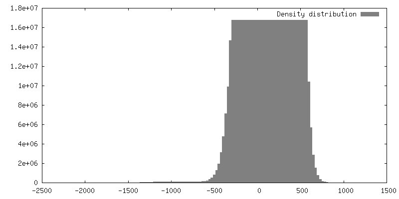
| 密度 |
| ||||||||||||||||||||||||||||||||
| 対称性 | 空間群: 1 | ||||||||||||||||||||||||||||||||
| 詳細 | EMDB XML:
|
 Z (Sec.)
 Y (Row.)
 X (Col.)


-添付データ
- 試料の構成要素
-全体 : HeLa Kyoto cell
| 全体 | 名称: HeLa Kyoto cell |
|---|---|
| 要素 |
|
-超分子 #1: HeLa Kyoto cell
| 超分子 | 名称: HeLa Kyoto cell / タイプ: cell / ID: 1 / 親要素: 0 |
|---|---|
| 由来(天然) | 生物種: Homo sapiens (ヒト) / 株: HeLa |
-実験情報
-構造解析
| 手法 | ネガティブ染色法, クライオ電子顕微鏡法 |
|---|---|
 解析 解析 | 電子線トモグラフィー法 |
| 試料の集合状態 | cell |
-試料調製
| 緩衝液 | pH: 7.4 詳細: DMEM without Riboflavin and Phenol Red, containing 10% FBS, 1% Pen/Strep, and 50 nM SiR-DNA |
|---|---|
| 染色 | タイプ: NEGATIVE / 材質: Uranyl acetate and lead citrate |
| 糖包埋 | 材質: Agar 100 Epoxy resin 詳細: Frozen cells were substituted in 0.1% uranyl acetate (UA), 2% Osmium tetroxide and 5% H2O in acetone following this temperature ramp: -90 C to -80 C for 10 hours, -80 C to -30 C for 10 hours, ...詳細: Frozen cells were substituted in 0.1% uranyl acetate (UA), 2% Osmium tetroxide and 5% H2O in acetone following this temperature ramp: -90 C to -80 C for 10 hours, -80 C to -30 C for 10 hours, -30 C for 4 hours, -30 C to 0 C for 6 hours, 0 C to 20 C for 4 hours, 20 C for 5-6 hours. |
| 凍結 | 凍結剤: NITROGEN |
| 加圧凍結法 | 装置: OTHER 詳細: High pressure freezing chamber was 1 mm thick, 6.0 mm diameter, with central cavities 5.0 mm x 5.0 mm x 25 um deep. The chamber had been in contact with 1-hexadecene.. The value given for _em_ ...詳細: High pressure freezing chamber was 1 mm thick, 6.0 mm diameter, with central cavities 5.0 mm x 5.0 mm x 25 um deep. The chamber had been in contact with 1-hexadecene.. The value given for _em_high_pressure_freezing.instrument is Leica EM ICE. This is not in a list of allowed values {'LEICA EM PACT2', 'BAL-TEC HPM 010', 'LEICA EM PACT', 'LEICA EM HPM100', 'OTHER', 'EMS-002 RAPID IMMERSION FREEZER'} so OTHER is written into the XML file. |
| Cryo protectant | 20% Ficoll-PM400 |
| 切片作成 | ウルトラミクロトーム - 装置: Leica Ultracut UCT / ウルトラミクロトーム - 温度: 298 K / ウルトラミクロトーム - 最終 厚さ: 250 |
| 位置合わせマーカー | Manufacturer: Cytodiagnostics / 直径: 15 nm |
- 電子顕微鏡法
| 顕微鏡 | FEI TECNAI F20 |
|---|---|
| 撮影 | フィルム・検出器のモデル: FEI EAGLE (4k x 4k) / 平均電子線量: 40.0 e/Å2 |
| 電子線 | 加速電圧: 200 kV / 電子線源:  FIELD EMISSION GUN FIELD EMISSION GUN |
| 電子光学系 | 照射モード: FLOOD BEAM / 撮影モード: BRIGHT FIELD / 最大 デフォーカス(公称値): 0.5 µm / 最小 デフォーカス(公称値): 0.2 µm |
| 実験機器 |  モデル: Tecnai F20 / 画像提供: FEI Company |
-画像解析
| 最終 再構成 | アルゴリズム: BACK PROJECTION / 使用した粒子像数: 110 |
|---|
