 Movie
Movie Controller

[English] 日本語
 Yorodumi
- EMDB-50025: Cryo-EM structure of the Pseudomonas aeruginosa PAO1 Type IV pilus -
+ Open data

- Basic information
| Entry |  | |||||||||||||||
|---|---|---|---|---|---|---|---|---|---|---|---|---|---|---|---|---|
| Title | Cryo-EM structure of the Pseudomonas aeruginosa PAO1 Type IV pilus | |||||||||||||||
 Map data Map data | ||||||||||||||||
 Sample Sample |
| |||||||||||||||
 Keywords Keywords | Type IV pilus / CELL ADHESION | |||||||||||||||
| Function / homology | Fimbrial protein pilin / Pilin (bacterial filament) / Prokaryotic N-terminal methylation motif / Prokaryotic N-terminal methylation site / Pilin-like / pilus / cell adhesion / Pilin Function and homology information Function and homology information | |||||||||||||||
| Biological species |   Pseudomonas aeruginosa (bacteria) Pseudomonas aeruginosa (bacteria) | |||||||||||||||
| Method | helical reconstruction / cryo EM / Resolution: 3.17 Å | |||||||||||||||
 Authors Authors | Ochner H / Boehning J / Wang Z / Tarafder A / Caspy I / Bharat TAM | |||||||||||||||
| Funding support |  United Kingdom, 4 items United Kingdom, 4 items
| |||||||||||||||
 Citation Citation | Journal: PLoS Pathog / Year: 2024 Title: Structure of the Pseudomonas aeruginosa PAO1 Type IV pilus. Authors: Hannah Ochner / Jan Böhning / Zhexin Wang / Abul K Tarafder / Ido Caspy / Tanmay A M Bharat / Abstract: Type IV pili (T4Ps) are abundant in many bacterial and archaeal species, where they play important roles in both surface sensing and twitching motility, with implications for adhesion, biofilm ...Type IV pili (T4Ps) are abundant in many bacterial and archaeal species, where they play important roles in both surface sensing and twitching motility, with implications for adhesion, biofilm formation and pathogenicity. While Type IV pilus (T4P) structures from other organisms have been previously solved, a high-resolution structure of the native, fully assembled T4P of Pseudomonas aeruginosa, a major human pathogen, would be valuable in a drug discovery context. Here, we report a 3.2 Å-resolution structure of the P. aeruginosa PAO1 T4P determined by electron cryomicroscopy (cryo-EM). PilA subunits constituting the T4P exhibit a classical pilin fold featuring an extended N-terminal α-helix linked to a C-terminal globular β-sheet-containing domain, which are packed tightly along the pilus, in line with models derived from previous cryo-EM data of the P. aeruginosa PAK strain. The N-terminal helices constitute the pilus core where they stabilise the tubular assembly via hydrophobic interactions. The α-helical core of the pilus is surrounded by the C-terminal globular domain of PilA that coats the outer surface of the pilus, mediating interactions with the surrounding environment. Comparison of the P. aeruginosa PAO1 T4P with T4P structures from other organisms, both at the level of the pilin subunits and the fully assembled pili, confirms previously described common architectural principles whilst highlighting key differences between members of this abundant class of prokaryotic filaments. This study provides a structural framework for understanding the molecular and cell biology of these important cellular appendages mediating interaction of prokaryotes to surfaces. | |||||||||||||||
| History |
|
- Structure visualization
| Supplemental images |
|---|

- Downloads & links
-EMDB archive
| Map data | emd_50025.map.gz | 228.7 MB | EMDB map data format | |
|---|---|---|---|---|
| Header (meta data) | emd-50025-v30.xmlemd-50025.xml | 15.3 KB 15.3 KB | Display Display | EMDB header |
| FSC (resolution estimation) | emd_50025_fsc.xml | 14.2 KB | Display | FSC data file |
| Images |  emd_50025.png emd_50025.png | 92.4 KB | ||
| Filedesc metadata | emd-50025.cif.gz | 5.3 KB | ||
| Others | emd_50025_half_map_1.map.gzemd_50025_half_map_2.map.gz | 194.1 MB 194.1 MB | ||
| Archive directory |  http://ftp.pdbj.org/pub/emdb/structures/EMD-50025ftp://ftp.pdbj.org/pub/emdb/structures/EMD-50025 http://ftp.pdbj.org/pub/emdb/structures/EMD-50025ftp://ftp.pdbj.org/pub/emdb/structures/EMD-50025 | HTTPS FTP |
-Related structure data
| Related structure data |  9ewxMC M: atomic model generated by this map C: citing same article ( |
|---|---|
| Similar structure data |
-Links
| EMDB pages | EMDB (EBI/PDBe) / EMDataResource |
|---|---|
| Related items in Molecule of the Month |

-Map
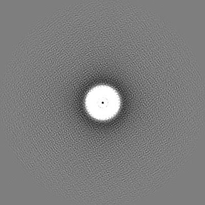
| File | Download / File: emd_50025.map.gz / Format: CCP4 / Size: 244.1 MB / Type: IMAGE STORED AS FLOATING POINT NUMBER (4 BYTES) | ||||||||||||||||||||||||||||||||||||
|---|---|---|---|---|---|---|---|---|---|---|---|---|---|---|---|---|---|---|---|---|---|---|---|---|---|---|---|---|---|---|---|---|---|---|---|---|---|
| Projections & slices | Image control
Images are generated by Spider. | ||||||||||||||||||||||||||||||||||||
| Voxel size | X=Y=Z: 0.824 Å | ||||||||||||||||||||||||||||||||||||
| Density |
| ||||||||||||||||||||||||||||||||||||
| Symmetry | Space group: 1 | ||||||||||||||||||||||||||||||||||||
| Details | EMDB XML:
|
 Z (Sec.)
 Y (Row.)
 X (Col.)


-Supplemental data
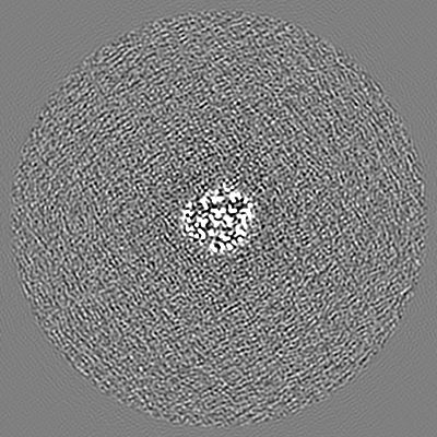
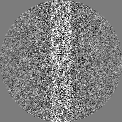
-Half map: #2
| File | emd_50025_half_map_1.map | ||||||||||||
|---|---|---|---|---|---|---|---|---|---|---|---|---|---|
| Projections & Slices |
| ||||||||||||
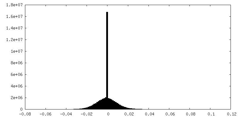
| Density Histograms |
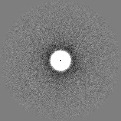


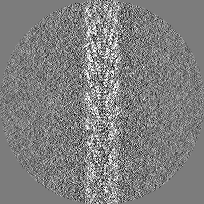


-Half map: #1
| File | emd_50025_half_map_2.map | ||||||||||||
|---|---|---|---|---|---|---|---|---|---|---|---|---|---|
| Projections & Slices |
| ||||||||||||
| Density Histograms |


- Sample components
-Entire : Pseudomonas aeruginosa PAO1 Type IV Pilus
| Entire | Name: Pseudomonas aeruginosa PAO1 Type IV Pilus |
|---|---|
| Components |
|
-Supramolecule #1: Pseudomonas aeruginosa PAO1 Type IV Pilus
| Supramolecule | Name: Pseudomonas aeruginosa PAO1 Type IV Pilus / type: complex / ID: 1 / Parent: 0 / Macromolecule list: all |
|---|---|
| Source (natural) | Organism: Pseudomonas aeruginosa (bacteria) |
| Molecular weight | Theoretical: 15 KDa |
-Macromolecule #1: Pilin
| Macromolecule | Name: Pilin / type: protein_or_peptide / ID: 1 / Number of copies: 23 / Enantiomer: LEVO |
|---|---|
| Source (natural) | Organism: Pseudomonas aeruginosa (bacteria) |
| Molecular weight | Theoretical: 14.878889 KDa |
| Sequence | String: FTLIELMIVV AIIGILAAIA IPQYQNYVAR SEGASALATI NPLKTTVEES LSRGIAGSKI KIGTTASTAT ETYVGVEPDA NKLGVIAVA IEDSGAGDIT FTFQTGTSSP KNATKVITLN RTADGVWACK STQDPMFTPK GCDN UniProtKB: Pilin |
-Experimental details
-Structure determination
| Method | cryo EM |
|---|---|
 Processing Processing | helical reconstruction |
| Aggregation state | helical array |
-Sample preparation
| Concentration | 6.46 mg/mL |
|---|---|
| Buffer | pH: 7.5 / Details: 50 mM Tris pH 7.5, 150 mM NaCl |
| Grid | Model: Quantifoil R3.5/1 / Material: COPPER/RHODIUM / Mesh: 200 / Support film - Material: CARBON / Support film - topology: HOLEY / Pretreatment - Type: GLOW DISCHARGE / Pretreatment - Time: 45 sec. |
| Vitrification | Cryogen name: ETHANE / Chamber humidity: 100 % / Chamber temperature: 283 K / Instrument: FEI VITROBOT MARK IV |
- Electron microscopy
| Microscope | TFS KRIOS |
|---|---|
| Image recording | Film or detector model: FEI FALCON IV (4k x 4k) / Number grids imaged: 1 / Number real images: 5679 / Average exposure time: 6.66 sec. / Average electron dose: 40.0 e/Å2 |
| Electron beam | Acceleration voltage: 300 kV / Electron source:  FIELD EMISSION GUN FIELD EMISSION GUN |
| Electron optics | C2 aperture diameter: 50.0 µm / Illumination mode: FLOOD BEAM / Imaging mode: BRIGHT FIELD / Nominal defocus max: 2.5 µm / Nominal defocus min: 1.0 µm |
| Experimental equipment |  Model: Titan Krios / Image courtesy: FEI Company |
-Image processing
| Final reconstruction | Applied symmetry - Helical parameters - Δz: 10.17 Å Applied symmetry - Helical parameters - Δ&Phi: 87.39 ° Applied symmetry - Helical parameters - Axial symmetry: C1 (asymmetric) Resolution.type: BY AUTHOR / Resolution: 3.17 Å / Resolution method: FSC 0.143 CUT-OFF / Software - Name: RELION (ver. 4.0) / Number images used: 114537 |
|---|---|
| Startup model | Type of model: OTHER / Details: Cylinder |
| Final angle assignment | Type: NOT APPLICABLE |
| FSC plot (resolution estimation) |  |
