 Movie
Movie Controller

[English] 日本語
 Yorodumi
- EMDB-50001: Structure of the peripheral stalk of the Polytomella ATPsynthase ... -
+ Open data

- Basic information
| Entry |  | |||||||||
|---|---|---|---|---|---|---|---|---|---|---|
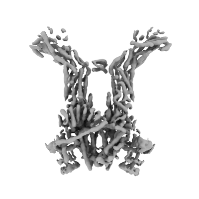
| Title | Structure of the peripheral stalk of the Polytomella ATPsynthase dimer in whole cells | |||||||||
 Map data Map data | ||||||||||
 Sample Sample |
| |||||||||
 Keywords Keywords | ATP synthesis / in situ / OXPHOS / MEMBRANE PROTEIN | |||||||||
| Biological species |  Polytomella sp. Pringsheim 198.80 (plant) Polytomella sp. Pringsheim 198.80 (plant) | |||||||||
| Method | subtomogram averaging / cryo EM / Resolution: 6.9 Å | |||||||||
 Authors Authors | Dietrich L / Kuehlbrandt W / Agip ANA | |||||||||
| Funding support |  Germany, 2 items Germany, 2 items
| |||||||||
 Citation Citation | Journal: Science / Year: 2024 Title: In situ structure and rotary states of mitochondrial ATP synthase in whole cells. Authors: Lea Dietrich / Ahmed-Noor A Agip / Christina Kunz / Andre Schwarz / Werner Kühlbrandt / Abstract: Cells depend on a continuous supply of adenosine triphosphate (ATP), the universal energy currency. In mitochondria, ATP is produced by a series of redox reactions, whereby an electrochemical ...Cells depend on a continuous supply of adenosine triphosphate (ATP), the universal energy currency. In mitochondria, ATP is produced by a series of redox reactions, whereby an electrochemical gradient is established across the inner mitochondrial membrane. The ATP synthase harnesses the energy of the gradient to generate ATP from adenosine diphosphate (ADP) and inorganic phosphate. We determined the structure of ATP synthase within mitochondria of the unicellular flagellate by electron cryo-tomography and subtomogram averaging at up to 4.2-angstrom resolution, revealing six rotary positions of the central stalk, subclassified into 21 substates of the F head. The ATP synthase forms helical arrays with multiple adjacent rows defining the cristae ridges. The structure of ATP synthase under native operating conditions in the presence of a membrane potential represents a pivotal step toward the analysis of membrane protein complexes in situ. | |||||||||
| History |
|
- Structure visualization
| Supplemental images |
|---|
- Downloads & links
-EMDB archive
| Map data | emd_50001.map.gz | 346.8 MB |  EMDB map data format EMDB map data format | |
|---|---|---|---|---|
| Header (meta data) | emd-50001-v30.xmlemd-50001.xml | 15.2 KB 15.2 KB | Display Display | EMDB header |
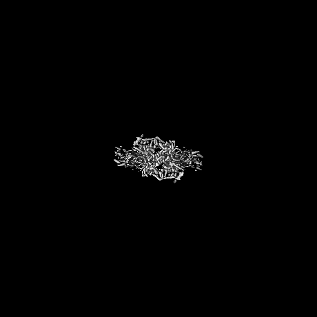
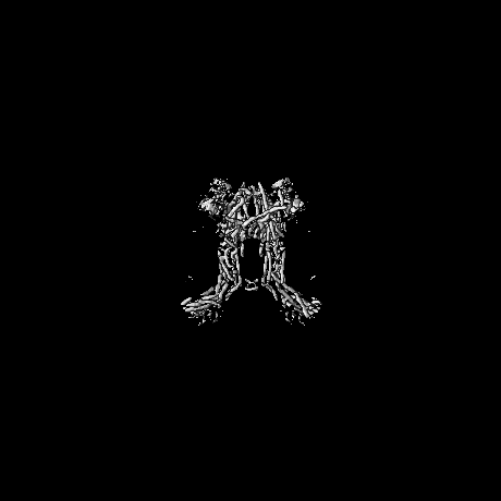
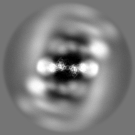
| Images |  emd_50001.png emd_50001.png | 63.2 KB | ||
| Filedesc metadata | emd-50001.cif.gz | 4.6 KB | ||
| Others | emd_50001_half_map_1.map.gzemd_50001_half_map_2.map.gz | 191 MB 191 MB | ||
| Archive directory |  http://ftp.pdbj.org/pub/emdb/structures/EMD-50001ftp://ftp.pdbj.org/pub/emdb/structures/EMD-50001 http://ftp.pdbj.org/pub/emdb/structures/EMD-50001ftp://ftp.pdbj.org/pub/emdb/structures/EMD-50001 | HTTPS FTP |
-Related structure data


-Links
| EMDB pages | EMDB (EBI/PDBe) / EMDataResource |
|---|
-Map
| File | Download / File: emd_50001.map.gz / Format: CCP4 / Size: 371.3 MB / Type: IMAGE STORED AS FLOATING POINT NUMBER (4 BYTES) | ||||||||||||||||||||||||||||||||||||
|---|---|---|---|---|---|---|---|---|---|---|---|---|---|---|---|---|---|---|---|---|---|---|---|---|---|---|---|---|---|---|---|---|---|---|---|---|---|
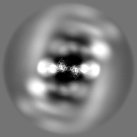
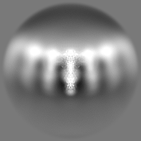
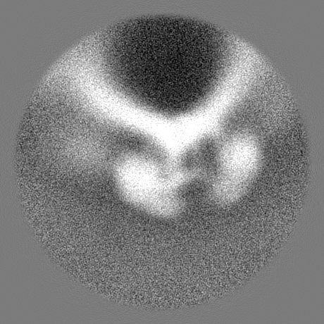
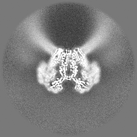
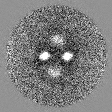
| Projections & slices | Image control
Images are generated by Spider. | ||||||||||||||||||||||||||||||||||||
| Voxel size | X=Y=Z: 1.63 Å | ||||||||||||||||||||||||||||||||||||
| Density |
| ||||||||||||||||||||||||||||||||||||
| Symmetry | Space group: 1 | ||||||||||||||||||||||||||||||||||||
| Details | EMDB XML:
|
 Z (Sec.)
 Y (Row.)
 X (Col.)


-Supplemental data
-Half map: #1
| File | emd_50001_half_map_1.map | ||||||||||||
|---|---|---|---|---|---|---|---|---|---|---|---|---|---|
| Projections & Slices |
| ||||||||||||
| Density Histograms |


-Half map: #2
| File | emd_50001_half_map_2.map | ||||||||||||
|---|---|---|---|---|---|---|---|---|---|---|---|---|---|
| Projections & Slices |
| ||||||||||||
| Density Histograms |


- Sample components
-Entire : Polytomella sp.
| Entire | Name: Polytomella sp. |
|---|---|
| Components |
|
-Supramolecule #1: Polytomella sp.
| Supramolecule | Name: Polytomella sp. / type: cell / ID: 1 / Parent: 0 |
|---|---|
| Source (natural) | Organism: Polytomella sp. Pringsheim 198.80 (plant) |
-Experimental details
-Structure determination
| Method | cryo EM |
|---|---|
 Processing Processing | subtomogram averaging |
| Aggregation state | cell |
-Sample preparation
| Buffer | pH: 7.4 |
|---|---|
| Grid | Model: Quantifoil R2/2 / Material: COPPER / Mesh: 200 / Support film - Material: CARBON / Support film - topology: HOLEY / Pretreatment - Type: GLOW DISCHARGE / Pretreatment - Time: 60 sec. |
| Vitrification | Cryogen name: ETHANE / Chamber humidity: 90 % / Chamber temperature: 298 K / Instrument: LEICA PLUNGER / Details: GP2. |
| Details | Liquid culture |
- Electron microscopy
| Microscope | FEI TITAN KRIOS |
|---|---|
| Specialist optics | Energy filter - Name: GIF Bioquantum / Energy filter - Slit width: 20 eV |
| Image recording | Film or detector model: GATAN K3 (6k x 4k) / Digitization - Dimensions - Width: 5760 pixel / Digitization - Dimensions - Height: 4040 pixel / Average electron dose: 1.9 e/Å2 |
| Electron beam | Acceleration voltage: 300 kV / Electron source:  FIELD EMISSION GUN FIELD EMISSION GUN |
| Electron optics | C2 aperture diameter: 70.0 µm / Illumination mode: FLOOD BEAM / Imaging mode: BRIGHT FIELD / Cs: 2.7 mm / Nominal defocus max: 4.5 µm / Nominal defocus min: 2.5 µm / Nominal magnification: 53000 |
| Sample stage | Specimen holder model: FEI TITAN KRIOS AUTOGRID HOLDER / Cooling holder cryogen: NITROGEN |
| Experimental equipment |  Model: Titan Krios / Image courtesy: FEI Company |
-Image processing
| Details | Image series was saved aligned and gain normalized |
|---|---|
| Final reconstruction | Applied symmetry - Point group: C2 (2 fold cyclic) / Resolution.type: BY AUTHOR / Resolution: 6.9 Å / Resolution method: FSC 0.143 CUT-OFF / Software - Name: Warp / Number subtomograms used: 238622 |
| Extraction | Number tomograms: 255 / Number images used: 363061 / Method: template matching / Software - Name: STOPGAP |
| Final angle assignment | Type: MAXIMUM LIKELIHOOD / Software - Name: RELION (ver. 3.1) |
