 Movie
Movie Controller


+ Open data

- Basic information
| Entry | 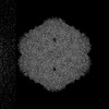 | |||||||||
|---|---|---|---|---|---|---|---|---|---|---|
| Title | Adeno-associated virus 9 variant AAV9.AAA.VQVGRTS | |||||||||
 Map data Map data | ||||||||||
 Sample Sample |
| |||||||||
 Keywords Keywords | parvovirus / virion / targeting / engineered / VIRUS LIKE PARTICLE | |||||||||
| Function / homology | Phospholipase A2-like domain / Phospholipase A2-like domain / Parvovirus coat protein VP2 / Parvovirus coat protein VP1/VP2 / Parvovirus coat protein VP1/VP2 / Capsid/spike protein, ssDNA virus / T=1 icosahedral viral capsid / structural molecule activity / Capsid protein VP1 Function and homology information Function and homology information | |||||||||
| Biological species |   Adeno-associated virus 9 Adeno-associated virus 9 | |||||||||
| Method | single particle reconstruction / cryo EM / Resolution: 1.84 Å | |||||||||
 Authors Authors | Choi H / Firnberg E / Tejada SKS / Yost SA / Giles AR / Liu Y / Danos O / Kaelber JT | |||||||||
| Funding support | 1 items
| |||||||||
 Citation Citation | Journal: To Be Published Title: Performance of low-voltage cryo-electron microscopes Authors: Choi H / Hou CFD / Firnberg E / Wei H / Petrou VI / Kaelber JT | |||||||||
| History |
|
- Structure visualization
| Supplemental images |
|---|

- Downloads & links
-EMDB archive
| Map data | emd_49965.map.gz | 777.5 MB | EMDB map data format | |
|---|---|---|---|---|
| Header (meta data) | emd-49965-v30.xmlemd-49965.xml | 21.2 KB 21.2 KB | Display Display | EMDB header |
| FSC (resolution estimation) | emd_49965_fsc.xml | 23.9 KB | Display | FSC data file |
| Images |  emd_49965.png emd_49965.png | 277.3 KB | ||
| Masks | emd_49965_msk_1.map | 824 MB | Mask map | |
| Filedesc metadata | emd-49965.cif.gz | 6.9 KB | ||
| Others | emd_49965_half_map_1.map.gzemd_49965_half_map_2.map.gz | 762.5 MB 762.5 MB | ||
| Archive directory |  http://ftp.pdbj.org/pub/emdb/structures/EMD-49965ftp://ftp.pdbj.org/pub/emdb/structures/EMD-49965 http://ftp.pdbj.org/pub/emdb/structures/EMD-49965ftp://ftp.pdbj.org/pub/emdb/structures/EMD-49965 | HTTPS FTP |
-Related structure data
| Related structure data |  9nzzMC  9o0eC  9o0fC M: atomic model generated by this map C: citing same article ( |
|---|---|
| Similar structure data |


-Links
| EMDB pages | EMDB (EBI/PDBe) / EMDataResource |
|---|---|
| Related items in Molecule of the Month |


-Map
| File | Download / File: emd_49965.map.gz / Format: CCP4 / Size: 824 MB / Type: IMAGE STORED AS FLOATING POINT NUMBER (4 BYTES) | ||||||||||||||||||||||||||||||||||||
|---|---|---|---|---|---|---|---|---|---|---|---|---|---|---|---|---|---|---|---|---|---|---|---|---|---|---|---|---|---|---|---|---|---|---|---|---|---|
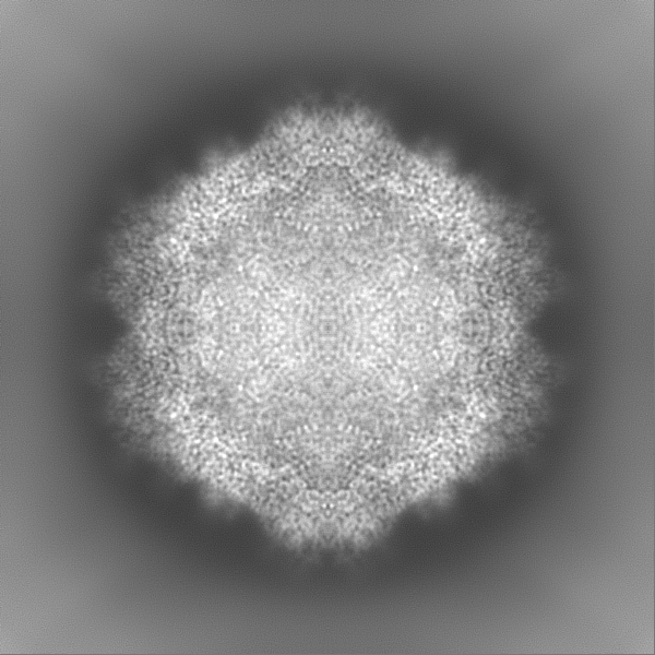
| Projections & slices | Image control
Images are generated by Spider. | ||||||||||||||||||||||||||||||||||||
| Voxel size | X=Y=Z: 0.628 Å | ||||||||||||||||||||||||||||||||||||
| Density |
| ||||||||||||||||||||||||||||||||||||
| Symmetry | Space group: 1 | ||||||||||||||||||||||||||||||||||||
| Details | EMDB XML:
|
 Z (Sec.)
 Y (Row.)
 X (Col.)


-Supplemental data
-Mask #1
| File | emd_49965_msk_1.map | ||||||||||||
|---|---|---|---|---|---|---|---|---|---|---|---|---|---|
| Projections & Slices |
| ||||||||||||
| Density Histograms |


-Half map: #2
| File | emd_49965_half_map_1.map | ||||||||||||
|---|---|---|---|---|---|---|---|---|---|---|---|---|---|
| Projections & Slices |
| ||||||||||||
| Density Histograms |


-Half map: #1
| File | emd_49965_half_map_2.map | ||||||||||||
|---|---|---|---|---|---|---|---|---|---|---|---|---|---|
| Projections & Slices |
| ||||||||||||
| Density Histograms |


- Sample components
-Entire : Adeno-associated virus 9
| Entire | Name: Adeno-associated virus 9 |
|---|---|
| Components |
|
-Supramolecule #1: Adeno-associated virus 9
| Supramolecule | Name: Adeno-associated virus 9 / type: virus / ID: 1 / Parent: 0 / Macromolecule list: #1 Details: NNN495-497AAA triple point mutation and insertion after S454 of VQVGRTS NCBI-ID: 235455 / Sci species name: Adeno-associated virus 9 / Sci species strain: AAA.NVG7 / Virus type: VIRUS-LIKE PARTICLE / Virus isolate: SEROTYPE / Virus enveloped: No / Virus empty: No |
|---|---|
| Host (natural) | Organism:  Homo sapiens (human) Homo sapiens (human) |
| Molecular weight | Theoretical: 5 MDa |
| Virus shell | Shell ID: 1 / Diameter: 226.0 Å / T number (triangulation number): 1 |
-Macromolecule #1: Capsid protein VP1
| Macromolecule | Name: Capsid protein VP1 / type: protein_or_peptide / ID: 1 / Number of copies: 1 / Enantiomer: LEVO |
|---|---|
| Source (natural) | Organism: Adeno-associated virus 9 / Strain: AAA.NVG7 |
| Molecular weight | Theoretical: 59.074168 KDa |
| Recombinant expression | Organism: Homo sapiens (human) |
| Sequence | String: DGVGSSSGNW HCDSQWLGDR VITTSTRTWA LPTYNNHLYK QISNSTSGGS SNDNAYFGYS TPWGYFDFNR FHCHFSPRDW QRLINNNWG FRPKRLNFKL FNIQVKEVTD NNGVKTIANN LTSTVQVFTD SDYQLPYVLG SAHEGCLPPF PADVFMIPQY G YLTLNDGS ...String: DGVGSSSGNW HCDSQWLGDR VITTSTRTWA LPTYNNHLYK QISNSTSGGS SNDNAYFGYS TPWGYFDFNR FHCHFSPRDW QRLINNNWG FRPKRLNFKL FNIQVKEVTD NNGVKTIANN LTSTVQVFTD SDYQLPYVLG SAHEGCLPPF PADVFMIPQY G YLTLNDGS QAVGRSSFYC LEYFPSQMLR TGNNFQFSYE FENVPFHSSY AHSQSLDRLM NPLIDQYLYY LSKTINGSVQ VG RTSGQNQ QTLKFSVAGP SNMAVQGRNY IPGPSYRQQR VSTTVTQAAA SEFAWPGASS WALNGRNSLM NPGPAMASHK EGE DRFFPL SGSLIFGKQG TGRDNVDADK VMITNEEEIK TTNPVATESY GQVATNHQSA QAQAQTGWVQ NQGILPGMVW QDRD VYLQG PIWAKIPHTD GNFHPSPLMG GFGMKHPPPQ ILIKNTPVPA DPPTAFNKDK LNSFITQYST GQVSVEIEWE LQKEN SKRW NPEIQYTSNY YKSNNVEFAV NTEGVYSEPR PIGTRYLTRN L UniProtKB: Capsid protein VP1 |
-Macromolecule #2: water
| Macromolecule | Name: water / type: ligand / ID: 2 / Number of copies: 584 / Formula: HOH |
|---|---|
| Molecular weight | Theoretical: 18.015 Da |
| Chemical component information |  ChemComp-HOH: |
-Experimental details
-Structure determination
| Method | cryo EM |
|---|---|
 Processing Processing | single particle reconstruction |
| Aggregation state | particle |
-Sample preparation
| Buffer | pH: 7.4 Component:
Details: PBS pH 7.4 | |||||||||||||||
|---|---|---|---|---|---|---|---|---|---|---|---|---|---|---|---|---|
| Grid | Model: UltrAuFoil R1.2/1.3 / Material: GOLD / Mesh: 300 / Support film - Material: GOLD / Support film - topology: HOLEY ARRAY / Pretreatment - Type: GLOW DISCHARGE / Pretreatment - Time: 300 sec. / Pretreatment - Atmosphere: AIR / Pretreatment - Pressure: 0.039 kPa | |||||||||||||||
| Vitrification | Cryogen name: ETHANE / Chamber humidity: 95 % / Chamber temperature: 298 K / Instrument: FEI VITROBOT MARK IV | |||||||||||||||
| Details | Concentration ~10^14 gc/mL |
- Electron microscopy
| Microscope | FEI TALOS ARCTICA |
|---|---|
| Image recording | Film or detector model: GATAN K2 QUANTUM (4k x 4k) / Detector mode: COUNTING / Digitization - Dimensions - Width: 3838 pixel / Digitization - Dimensions - Height: 3710 pixel / Digitization - Frames/image: 1-40 / Number grids imaged: 1 / Number real images: 2685 / Average exposure time: 2.0 sec. / Average electron dose: 28.36 e/Å2 |
| Electron beam | Acceleration voltage: 200 kV / Electron source:  FIELD EMISSION GUN FIELD EMISSION GUN |
| Electron optics | C2 aperture diameter: 50.0 µm / Calibrated defocus max: 1.7 µm / Calibrated defocus min: 0.4 µm / Illumination mode: FLOOD BEAM / Imaging mode: BRIGHT FIELD / Cs: 2.7 mm / Nominal defocus max: 1.5 µm / Nominal defocus min: 0.5 µm / Nominal magnification: 130000 |
| Sample stage | Specimen holder model: FEI TITAN KRIOS AUTOGRID HOLDER / Cooling holder cryogen: NITROGEN |
| Experimental equipment |  Model: Talos Arctica / Image courtesy: FEI Company |


