[English] 日本語
Yorodumi
- EMDB-49932: Cryo-EM structure of the glycosyltransferase GtrB in the apo stat... -

+
Open data


ID or keywords:

Loading...

-
Basic information

Entry
Database: EMDB / ID: EMD-49932
TitleCryo-EM structure of the glycosyltransferase GtrB in the apo state (octamer volume)
Map datatructure of the glycosyltransferase GtrB in the apo state (octamer volume)
Sample
  • Complex: Apo-GtrB
    • Protein or peptide: Glycosyltransferase sll0501
Keywordsglycosyltransferase / polyisoprenyl / MEMBRANE PROTEIN
Function / homologyTransferases; Glycosyltransferases / : / Glycosyltransferase 2-like / Glycosyl transferase family 2 / glycosyltransferase activity / Nucleotide-diphospho-sugar transferases / plasma membrane / Uncharacterized glycosyltransferase sll0501
Function and homology information
Biological speciesSynechocystis sp. PCC 6803 substr. Kazusa (bacteria)
Methodsingle particle reconstruction / cryo EM / Resolution: 2.79 Å
AuthorsMorgan RT / Motta S / Gil-Iturbe E / di Muccio G / Bhattacharjee B / Romagnoli A / Anwar MT / Mishra B / Ashraf K / Bang I ...Morgan RT / Motta S / Gil-Iturbe E / di Muccio G / Bhattacharjee B / Romagnoli A / Anwar MT / Mishra B / Ashraf K / Bang I / di Marino D / Lowary TL / Quick M / Petrou VI / Stowell MHB / Nygaard R / Mancia F
Funding support United States, 5 items
OrganizationGrant numberCountry
National Institutes of Health/National Institute of General Medical Sciences (NIH/NIGMS)R35GM132120 United States
National Institutes of Health/National Institute of Neurological Disorders and Stroke (NIH/NINDS)R01NS120496 United States
National Institutes of Health/National Institute of General Medical Sciences (NIH/NIGMS)K99GM123228 United States
National Institutes of Health/National Institute of General Medical Sciences (NIH/NIGMS)R00GM123228 United States
National Institutes of Health/National Institute of General Medical Sciences (NIH/NIGMS)R35GM150831 United States
Citation
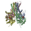
Journal: Nat Commun / Year: 2025
Title: Mechanistic snapshots of lipid-linked sugar transfer.
Authors: Ryan T Morgan / Stefano Motta / Eva Gil-Iturbe / Biddut Bhattacharjee / Mohammad T Anwar / Giovanni Di Muccio / Alice Romagnoli / Bedangshu Mishra / Khuram U Ashraf / Injin Bang / Daniele Di ...Authors: Ryan T Morgan / Stefano Motta / Eva Gil-Iturbe / Biddut Bhattacharjee / Mohammad T Anwar / Giovanni Di Muccio / Alice Romagnoli / Bedangshu Mishra / Khuram U Ashraf / Injin Bang / Daniele Di Marino / Todd L Lowary / Matthias Quick / Vasileios I Petrou / Michael H B Stowell / Rie Nygaard / Filippo Mancia /
Abstract: Enzymes undergo dynamic conformational changes during catalysis, yet conventional high-resolution structural methods typically capture only the most stable states. Here, we address this gap using ...Enzymes undergo dynamic conformational changes during catalysis, yet conventional high-resolution structural methods typically capture only the most stable states. Here, we address this gap using rapid UV photolysis of a chemically caged substrate with cryogenic time-resolved electron microscopy (cryo-TREM). We elucidate the catalytic mechanism of GtrB, a membrane-bound glycosyltransferase that transfers glucose from UDP-glucose to the lipid carrier undecaprenyl phosphate. We visualized how GtrB, which has an active site ~15 Å from the membrane, transitions during the catalytic cycle to move each substrate in proximity for catalysis. From a single dataset, we resolved distinct conformational states: the initial substrate-bound state, a catalytically poised intermediate, and the product-bound state. Through molecular dynamics simulations and biochemical analyses, we identify coordinated movements within the active site that drive catalysis. These findings provide a molecular framework for understanding how glycosyltransferases function and highlight a broadly applicable strategy for capturing dynamic enzymatic states in native-like environments.
History
DepositionMar 27, 2025-
Header (metadata) releaseDec 17, 2025-
Map releaseDec 17, 2025-
UpdateDec 24, 2025-
Current statusDec 24, 2025Processing site: RCSB / Status: Released

-
Structure visualization

Supplemental images

Downloads & links

-
Map

FileDownload / File: emd_49932.map.gz / Format: CCP4 / Size: 103 MB / Type: IMAGE STORED AS FLOATING POINT NUMBER (4 BYTES)
Annotationtructure of the glycosyltransferase GtrB in the apo state (octamer volume)
Projections & slices

Image control

Size
Brightness
Contrast
Others
AxesZ (Sec.)Y (Row.)X (Col.)
0.95 Å/pix.
x 300 pix.
= 285. Å
0.95 Å/pix.
x 300 pix.
= 285. Å
0.95 Å/pix.
x 300 pix.
= 285. Å

Surface

Projections

Slices (1/3)

Slices (1/2)

Slices (2/3)

Images are generated by Spider.

Voxel sizeX=Y=Z: 0.95 Å
Density
Contour LevelBy AUTHOR: 0.11
Minimum - Maximum-1.122666 - 1.321182
Average (Standard dev.)0.0005570777 (±0.034025818)
SymmetrySpace group: 1
Details

EMDB XML:

Map geometry
Axis orderXYZ
Origin000
Dimensions300300300
Spacing300300300
CellA=B=C: 285.0 Å
α=β=γ: 90.0 °

-
Supplemental data

-
Mask #1

Fileemd_49932_msk_1.map
Projections & Slices
AxesZYX

Projections

Slices (1/2)
Density Histograms

-
Half map: #2

Fileemd_49932_half_map_1.map
Projections & Slices
AxesZYX

Projections

Slices (1/2)
Density Histograms

-
Half map: #1

Fileemd_49932_half_map_2.map
Projections & Slices
AxesZYX

Projections

Slices (1/2)
Density Histograms

-
Sample components

-
Entire : Apo-GtrB

EntireName: Apo-GtrB
Components
  • Complex: Apo-GtrB
    • Protein or peptide: Glycosyltransferase sll0501

-
Supramolecule #1: Apo-GtrB

SupramoleculeName: Apo-GtrB / type: complex / ID: 1 / Parent: 0 / Macromolecule list: all
Source (natural)Organism: Synechocystis sp. PCC 6803 substr. Kazusa (bacteria)
Molecular weightTheoretical: 293.346 KDa

-
Macromolecule #1: Glycosyltransferase sll0501

MacromoleculeName: Glycosyltransferase sll0501 / type: protein_or_peptide / ID: 1 / Number of copies: 8 / Enantiomer: LEVO / EC number: Transferases; Glycosyltransferases
Source (natural)Organism: Synechocystis sp. PCC 6803 substr. Kazusa (bacteria)
Molecular weightTheoretical: 39.872016 KDa
Recombinant expressionOrganism: Escherichia coli 'BL21-Gold(DE3)pLysS AG' (bacteria)
SequenceString: MHHHHHHHHH HSSGVDLGTE NLYFQSNATI ELSIVIPMYN EEDNLEHLFA RLLEVLTPLK ITYEIICVND GSKDKTLKQL IDCYQSNRQ IKIVNLSRNF GKEIALSAGI DYAQGNAVIP IDADLQDPPE LIHELVDKWR EGYDIVYATR RSRQGETWVK Q FTAKMFYK ...String:
MHHHHHHHHH HSSGVDLGTE NLYFQSNATI ELSIVIPMYN EEDNLEHLFA RLLEVLTPLK ITYEIICVND GSKDKTLKQL IDCYQSNRQ IKIVNLSRNF GKEIALSAGI DYAQGNAVIP IDADLQDPPE LIHELVDKWR EGYDIVYATR RSRQGETWVK Q FTAKMFYK VIGRMTEIKI PPNTGDFRLM DRKVVNAIKQ LPERTRFMKG LFAWVGYRQT FVLFDREPRF QGQTKWNYWK LW NFALDGI FSFSLLPLKV WTYLGSIISL LSLAYASFLI LKTITLGVDV PGYASLMVAI LFLGGVQLIS LGVIGEYLGR VYE EVKARP LYLVSDLWGL EYLPLEKLN

UniProtKB: Uncharacterized glycosyltransferase sll0501

-
Experimental details

-
Structure determination

Methodcryo EM
Processingsingle particle reconstruction
Aggregation stateparticle

-
Sample preparation

Concentration1.2 mg/mL
BufferpH: 7.5
VitrificationCryogen name: ETHANE / Chamber humidity: 100 % / Chamber temperature: 277.15 K / Instrument: FEI VITROBOT MARK IV

-
Electron microscopy

MicroscopeFEI POLARA 300
Image recordingFilm or detector model: GATAN K3 BIOQUANTUM (6k x 4k) / Number grids imaged: 1 / Number real images: 3757 / Average exposure time: 4.0 sec. / Average electron dose: 70.91 e/Å2
Electron beamAcceleration voltage: 300 kV / Electron source: FIELD EMISSION GUN
Electron opticsCalibrated defocus max: 2.5 µm / Calibrated defocus min: 1.5 µm / Illumination mode: FLOOD BEAM / Imaging mode: BRIGHT FIELD / Nominal defocus max: 2.4 µm / Nominal defocus min: 1.0 µm / Nominal magnification: 105000
Experimental equipment
Model: Tecnai Polara / Image courtesy: FEI Company

+
Image processing

CTF correctionType: PHASE FLIPPING AND AMPLITUDE CORRECTION
Startup modelType of model: PDB ENTRY
PDB model - PDB ID:
Final reconstructionResolution.type: BY AUTHOR / Resolution: 2.79 Å / Resolution method: FSC 0.143 CUT-OFF / Number images used: 152605
Initial angle assignmentType: MAXIMUM LIKELIHOOD
Final angle assignmentType: MAXIMUM LIKELIHOOD
FSC plot (resolution estimation)

+
About Yorodumi

-
News

-
Feb 9, 2022. New format data for meta-information of EMDB entries

New format data for meta-information of EMDB entries

  • Version 3 of the EMDB header file is now the official format.
  • The previous official version 1.9 will be removed from the archive.

Related info.:EMDB header

External links:wwPDB to switch to version 3 of the EMDB data model

-
Aug 12, 2020. Covid-19 info

Covid-19 info

URL: https://pdbj.org/emnavi/covid19.php

New page: Covid-19 featured information page in EM Navigator.

Related info.:Covid-19 info / Mar 5, 2020. Novel coronavirus structure data

+
Mar 5, 2020. Novel coronavirus structure data

Novel coronavirus structure data

Related info.:Yorodumi Speices / Aug 12, 2020. Covid-19 info

External links:COVID-19 featured content - PDBj / Molecule of the Month (242):Coronavirus Proteases

+
Jan 31, 2019. EMDB accession codes are about to change! (news from PDBe EMDB page)

EMDB accession codes are about to change! (news from PDBe EMDB page)

  • The allocation of 4 digits for EMDB accession codes will soon come to an end. Whilst these codes will remain in use, new EMDB accession codes will include an additional digit and will expand incrementally as the available range of codes is exhausted. The current 4-digit format prefixed with “EMD-” (i.e. EMD-XXXX) will advance to a 5-digit format (i.e. EMD-XXXXX), and so on. It is currently estimated that the 4-digit codes will be depleted around Spring 2019, at which point the 5-digit format will come into force.
  • The EM Navigator/Yorodumi systems omit the EMD- prefix.

Related info.:Q: What is EMD? / ID/Accession-code notation in Yorodumi/EM Navigator

External links:EMDB Accession Codes are Changing Soon! / Contact to PDBj

+
Jul 12, 2017. Major update of PDB

Major update of PDB

  • wwPDB released updated PDB data conforming to the new PDBx/mmCIF dictionary.
  • This is a major update changing the version number from 4 to 5, and with Remediation, in which all the entries are updated.
  • In this update, many items about electron microscopy experimental information are reorganized (e.g. em_software).
  • Now, EM Navigator and Yorodumi are based on the updated data.

External links:wwPDB Remediation / Enriched Model Files Conforming to OneDep Data Standards Now Available in the PDB FTP Archive

-
Yorodumi

Thousand views of thousand structures

  • Yorodumi is a browser for structure data from EMDB, PDB, SASBDB, etc.
  • This page is also the successor to EM Navigator detail page, and also detail information page/front-end page for Omokage search.
  • The word "yorodu" (or yorozu) is an old Japanese word meaning "ten thousand". "mi" (miru) is to see.

Related info.:EMDB / PDB / SASBDB / Comparison of 3 databanks / Yorodumi Search / Aug 31, 2016. New EM Navigator & Yorodumi / Yorodumi Papers / Jmol/JSmol / Function and homology information / Changes in new EM Navigator and Yorodumi

Read more