National Institutes of Health/National Institute Of Allergy and Infectious Diseases (NIH/NIAID)
United States
Citation
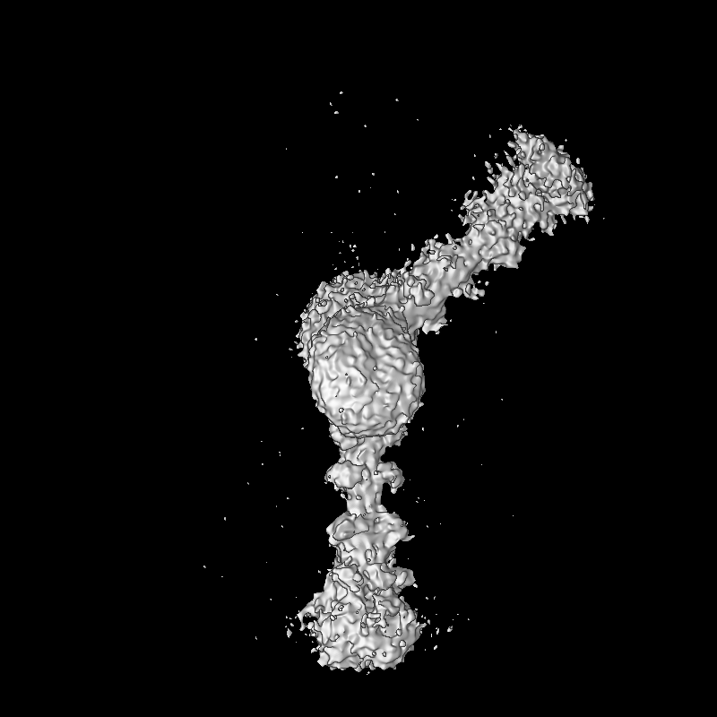
Journal: Nat Commun / Year: 2025 Title: Effect of the S2' site cleavage on SARS-CoV-2 spike. Authors: Wei Shi / G M Jonaid / Md Golam Kibria / Jacob Allen / Hanqin Peng / Sophia Rits-Volloch / Haisun Zhu / Shaowei Wang / Richard M Walsh / Jianming Lu / Bing Chen / Abstract: SARS-CoV-2 initiates infection of host cells by fusing its envelope lipid bilayer with the cell membrane. To overcome kinetic barriers for membrane fusion, the virus-encoded spike (S) protein refolds ...SARS-CoV-2 initiates infection of host cells by fusing its envelope lipid bilayer with the cell membrane. To overcome kinetic barriers for membrane fusion, the virus-encoded spike (S) protein refolds from a metastable prefusion state to a lower energy, stable postfusion conformation. The protein is first split into S1 and S2 fragments at a proteolytic site after synthesis, and presumably further cleaved at a second site, known as the S2' site, before membrane fusion can occur. Here, we report a cryo-EM structure of S2 fragment after the S2' cleavage, possibly representing a late fusion intermediate conformation, in which the fusion peptide and transmembrane segment have yet to pack together, distinct from the final, postfusion state. Functional assays demonstrate that the S2' cleavage accelerates membrane fusion, probably by stabilizing membrane fusion intermediates. These results advance our understanding of SARS-CoV-2 entry and may guide intervention strategies against pathogenetic coronaviruses.
In the structure databanks used in Yorodumi, some data are registered as the other names, "COVID-19 virus" and "2019-nCoV". Here are the details of the virus and the list of structure data.
Jan 31, 2019. EMDB accession codes are about to change! (news from PDBe EMDB page)
EMDB accession codes are about to change! (news from PDBe EMDB page)
The allocation of 4 digits for EMDB accession codes will soon come to an end. Whilst these codes will remain in use, new EMDB accession codes will include an additional digit and will expand incrementally as the available range of codes is exhausted. The current 4-digit format prefixed with “EMD-” (i.e. EMD-XXXX) will advance to a 5-digit format (i.e. EMD-XXXXX), and so on. It is currently estimated that the 4-digit codes will be depleted around Spring 2019, at which point the 5-digit format will come into force.
The EM Navigator/Yorodumi systems omit the EMD- prefix.
Related info.:Q: What is EMD? / ID/Accession-code notation in Yorodumi/EM Navigator
Yorodumi is a browser for structure data from EMDB, PDB, SASBDB, etc.
This page is also the successor to EM Navigator detail page, and also detail information page/front-end page for Omokage search.
The word "yorodu" (or yorozu) is an old Japanese word meaning "ten thousand". "mi" (miru) is to see.
Related info.:EMDB / PDB / SASBDB / Comparison of 3 databanks / Yorodumi Search / Aug 31, 2016. New EM Navigator & Yorodumi / Yorodumi Papers / Jmol/JSmol / Function and homology information / Changes in new EM Navigator and Yorodumi
 Movie
Movie Controller

 Yorodumi
 Open data
 Basic information
 Map data
 Sample
 Keywords
 Severe acute respiratory syndrome coronavirus 2
 Authors
 United States, 1 items
  Citation
 Structure visualization
 Downloads & links
 EMDB map data format
 emd_49921.png
 http://ftp.pdbj.org/pub/emdb/structures/EMD-49921


 Z (Sec.)
 Y (Row.)
 X (Col.)


 Sample components
 Processing
 Electron microscopy
 FIELD EMISSION GUN

