[English] 日本語
Yorodumi
- EMDB-49589: A membrane protein with cofactor determined by single-particle CryoEM -

+
Open data


ID or keywords:

Loading...

-
Basic information

Entry
Database: EMDB / ID: EMD-49589
TitleA membrane protein with cofactor determined by single-particle CryoEM
Map datasharpened map for opsin with mb7 (megabody 7)
Sample
  • Complex: Opsin with Mb7 (Megabody 7)
    • Protein or peptide: Megabody 7
    • Protein or peptide: Rhodopsin
Keywordsopsin / membrane protein / megabody / nanobody
Function / homology
Function and homology information


Opsins / VxPx cargo-targeting to cilium / rod bipolar cell differentiation / sperm head plasma membrane / absorption of visible light / opsin binding / The canonical retinoid cycle in rods (twilight vision) / G protein-coupled opsin signaling pathway / 11-cis retinal binding / podosome assembly ...Opsins / VxPx cargo-targeting to cilium / rod bipolar cell differentiation / sperm head plasma membrane / absorption of visible light / opsin binding / The canonical retinoid cycle in rods (twilight vision) / G protein-coupled opsin signaling pathway / 11-cis retinal binding / podosome assembly / G protein-coupled photoreceptor activity / photoreceptor inner segment membrane / cellular response to light stimulus / rod photoreceptor outer segment / G protein-coupled receptor complex / Inactivation, recovery and regulation of the phototransduction cascade / thermotaxis / Activation of the phototransduction cascade / outer membrane / detection of temperature stimulus involved in thermoception / response to light intensity / photoreceptor cell maintenance / arrestin family protein binding / photoreceptor outer segment membrane / G alpha (i) signalling events / response to light stimulus / phototransduction, visible light / phototransduction / G-protein alpha-subunit binding / photoreceptor outer segment / visual perception / guanyl-nucleotide exchange factor activity / microtubule cytoskeleton organization / cell-cell junction / photoreceptor disc membrane / sperm midpiece / gene expression / G protein-coupled receptor signaling pathway / Golgi membrane / zinc ion binding / identical protein binding / membrane / plasma membrane
Similarity search - Function
Rhodopsin, N-terminal / Amino terminal of the G-protein receptor rhodopsin / Rhodopsin / Opsin / Visual pigments (opsins) retinal binding site / Visual pigments (opsins) retinal binding site. / : / Serpentine type 7TM GPCR chemoreceptor Srsx / G-protein coupled receptors family 1 signature. / 7 transmembrane receptor (rhodopsin family) ...Rhodopsin, N-terminal / Amino terminal of the G-protein receptor rhodopsin / Rhodopsin / Opsin / Visual pigments (opsins) retinal binding site / Visual pigments (opsins) retinal binding site. / : / Serpentine type 7TM GPCR chemoreceptor Srsx / G-protein coupled receptors family 1 signature. / 7 transmembrane receptor (rhodopsin family) / G protein-coupled receptor, rhodopsin-like / GPCR, rhodopsin-like, 7TM / G-protein coupled receptors family 1 profile.
Similarity search - Domain/homology
Biological speciesBos taurus (domestic cattle) / synthetic construct (others)
Methodsingle particle reconstruction / cryo EM / Resolution: 3.94 Å
AuthorsSuder DS / Gonen S
Funding support United States, 1 items
OrganizationGrant numberCountry
National Institutes of Health/National Institute of General Medical Sciences (NIH/NIGMS)R35-GM142797 United States
Citation
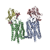
Journal: Proc Natl Acad Sci U S A / Year: 2026
Title: Structural analysis of rhodopsin states in megabody complexes.
Authors: David Salom / Diana S Suder / Wei Huang / Arum Wu / Els Pardon / Jan Steyaert / Philip D Kiser / Derek J Taylor / Shane Gonen / Krzysztof Palczewski /
Abstract: Rhodopsin, the most intensively studied G protein-coupled receptor (GPCR), is activated by light-induced isomerization of its chromophore 11--retinal. This study employed cryogenic electron ...Rhodopsin, the most intensively studied G protein-coupled receptor (GPCR), is activated by light-induced isomerization of its chromophore 11--retinal. This study employed cryogenic electron microscopy (cryo-EM) to investigate rhodopsin structure using a megabody (Mb7) as a negative allosteric modulator. Three distinct cryo-EM structures were solved: ground-state rhodopsin, photoactivated rhodopsin, and apo-rhodopsin, all in complex with Mb7. Photoactivated rhodopsin and apo-rhodopsin, both in complex with Mb7, maintain a conformation remarkably similar to ground-state rhodopsin rather than adopting a Meta-II-like conformation. Structural elements, including the conserved residues of the NPxxY motif and the ionic lock, remain in positions corresponding to inactive rhodopsin. The megabody forms extensive interactions with rhodopsin's extracellular loop 2, N terminus, and glycans. The findings demonstrate that Mb7 stabilizes photoactivated rhodopsin in a Meta-I-like conformation, preventing progression to the active Meta-II state through specific immobilization of the extracellular domain. This work establishes a foundation for cryo-EM-guided discovery of ligands modulating rhodopsin.
History
DepositionMar 6, 2025-
Header (metadata) releaseFeb 18, 2026-
Map releaseFeb 18, 2026-
UpdateFeb 18, 2026-
Current statusFeb 18, 2026Processing site: RCSB / Status: Released

-
Structure visualization

Supplemental images

Downloads & links

-
Map

FileDownload / File: emd_49589.map.gz / Format: CCP4 / Size: 103 MB / Type: IMAGE STORED AS FLOATING POINT NUMBER (4 BYTES)
Annotationsharpened map for opsin with mb7 (megabody 7)
Projections & slices

Image control

Size
Brightness
Contrast
Others
AxesZ (Sec.)Y (Row.)X (Col.)
0.74 Å/pix.
x 300 pix.
= 222.9 Å
0.74 Å/pix.
x 300 pix.
= 222.9 Å
0.74 Å/pix.
x 300 pix.
= 222.9 Å

Surface

Projections

Slices (1/3)

Slices (1/2)

Slices (2/3)

Images are generated by Spider.

Voxel sizeX=Y=Z: 0.743 Å
Density
Contour LevelBy AUTHOR: 0.2
Minimum - Maximum-0.38813388 - 0.6536589
Average (Standard dev.)0.000472545 (±0.014357131)
SymmetrySpace group: 1
Details

EMDB XML:

Map geometry
Axis orderXYZ
Origin000
Dimensions300300300
Spacing300300300
CellA=B=C: 222.9 Å
α=β=γ: 90.0 °

-
Supplemental data

-
Mask #1

Fileemd_49589_msk_1.map
Projections & Slices
AxesZYX

Projections

Slices (1/2)
Density Histograms

-
Mask #2

Fileemd_49589_msk_2.map
Projections & Slices
AxesZYX

Projections

Slices (1/2)
Density Histograms

-
Half map: resampled coordinates for half map B as requested...

Fileemd_49589_half_map_1.map
Annotationresampled coordinates for half map B as requested to match primary map. half map is unsharpened.
Projections & Slices
AxesZYX

Projections

Slices (1/2)
Density Histograms

-
Half map: resampled coordinates for half map A as requested...

Fileemd_49589_half_map_2.map
Annotationresampled coordinates for half map A as requested to match primary map. half map is unsharpened.
Projections & Slices
AxesZYX

Projections

Slices (1/2)
Density Histograms

-
Sample components

-
Entire : Opsin with Mb7 (Megabody 7)

EntireName: Opsin with Mb7 (Megabody 7)
Components
  • Complex: Opsin with Mb7 (Megabody 7)
    • Protein or peptide: Megabody 7
    • Protein or peptide: Rhodopsin

-
Supramolecule #1: Opsin with Mb7 (Megabody 7)

SupramoleculeName: Opsin with Mb7 (Megabody 7) / type: complex / ID: 1 / Parent: 0 / Macromolecule list: all
Source (natural)Organism: Bos taurus (domestic cattle)

-
Macromolecule #1: Megabody 7

MacromoleculeName: Megabody 7 / type: protein_or_peptide / ID: 1 / Number of copies: 1 / Enantiomer: LEVO
Source (natural)Organism: synthetic construct (others)
Molecular weightTheoretical: 55.284621 KDa
Recombinant expressionOrganism: Escherichia coli (E. coli)
SequenceString: QVQLVESGGG LVQTKTTTSV IDTTNDAQNL LTQAQTIVNT LKDYCPILIA KSSSSNGGTN NANTPSWQTA GGGKNSCATF GAEFSAASD MINNAQKIVQ ETQQLSANQP KNITQPHNLN LNSPSSLTAL AQKMLKNAQS QAEILKLANQ VESDFNKLSS G HLKDYIGK ...String:
QVQLVESGGG LVQTKTTTSV IDTTNDAQNL LTQAQTIVNT LKDYCPILIA KSSSSNGGTN NANTPSWQTA GGGKNSCATF GAEFSAASD MINNAQKIVQ ETQQLSANQP KNITQPHNLN LNSPSSLTAL AQKMLKNAQS QAEILKLANQ VESDFNKLSS G HLKDYIGK CDASAISSAN MTMQNQKNNW GNGCAGVEET QSLLKTSAAD FNNQTPQINQ AQNLANTLIQ ELGNNTYEQL SR LLTNDNG TNSKTSAQAI NQAVNNLNER AKTLAGGTTN SPAYQATLLA LRSVLGLWNS MGYAVICGGY TKSPGENNQK DFH YTDENG NGTTINCGGS TNSNGTHSYN GTNTLKADKN VSLSIEQYEK IHEAYQILSK ALKQAGLAPL NSKGEKLEAH VTTS KYGSL RLSCAASGFT FSRYVMNWVR QAPGKGLEWV SGISRDGSYT DYADSVKGRF TISRDNAKDT LYLQMNSLKP EDTAV YYCS KYNSVVTTPP GQGTQVTVSS HHHHHHEPEA

-
Macromolecule #2: Rhodopsin

MacromoleculeName: Rhodopsin / type: protein_or_peptide / ID: 2 / Number of copies: 1 / Enantiomer: LEVO
Source (natural)Organism: Bos taurus (domestic cattle)
Molecular weightTheoretical: 39.031457 KDa
SequenceString: MNGTEGPNFY VPFSNKTGVV RSPFEAPQYY LAEPWQFSML AAYMFLLIML GFPINFLTLY VTVQHKKLRT PLNYILLNLA VADLFMVFG GFTTTLYTSL HGYFVFGPTG CNLEGFFATL GGEIALWSLV VLAIERYVVV CKPMSNFRFG ENHAIMGVAF T WVMALACA ...String:
MNGTEGPNFY VPFSNKTGVV RSPFEAPQYY LAEPWQFSML AAYMFLLIML GFPINFLTLY VTVQHKKLRT PLNYILLNLA VADLFMVFG GFTTTLYTSL HGYFVFGPTG CNLEGFFATL GGEIALWSLV VLAIERYVVV CKPMSNFRFG ENHAIMGVAF T WVMALACA APPLVGWSRY IPEGMQCSCG IDYYTPHEET NNESFVIYMF VVHFIIPLIV IFFCYGQLVF TVKEAAAQQQ ES ATTQKAE KEVTRMVIIM VIAFLICWLP YAGVAFYIFT HQGSDFGPIF MTIPAFFAKT SAVYNPVIYI MMNKQFRNCM VTT LCCGKN PLGDDEASTT VSKTETSQVA PA

UniProtKB: Rhodopsin

-
Experimental details

-
Structure determination

Methodcryo EM
Processingsingle particle reconstruction
Aggregation stateparticle

-
Sample preparation

BufferpH: 7.4
GridModel: Quantifoil R1.2/1.3 / Material: GOLD / Mesh: 300 / Pretreatment - Type: GLOW DISCHARGE / Pretreatment - Time: 45 sec.
VitrificationCryogen name: ETHANE / Details: LEICA EM GP2.

-
Electron microscopy

MicroscopeTFS KRIOS
Image recordingFilm or detector model: FEI FALCON IV (4k x 4k) / Average electron dose: 70.0 e/Å2
Electron beamAcceleration voltage: 300 kV / Electron source: FIELD EMISSION GUN
Electron opticsIllumination mode: FLOOD BEAM / Imaging mode: BRIGHT FIELD / Nominal defocus max: 2.5 µm / Nominal defocus min: 0.8 µm
Experimental equipment
Model: Titan Krios / Image courtesy: FEI Company

+
Image processing

CTF correctionType: PHASE FLIPPING AND AMPLITUDE CORRECTION
Startup modelType of model: OTHER
Details: ab initio model from subsample of particles from this dataset
Final reconstructionApplied symmetry - Point group: C1 (asymmetric) / Resolution.type: BY AUTHOR / Resolution: 3.94 Å / Resolution method: FSC 0.143 CUT-OFF / Software - Name: cryoSPARC / Number images used: 337101
Initial angle assignmentType: MAXIMUM LIKELIHOOD
Final angle assignmentType: MAXIMUM LIKELIHOOD
FSC plot (resolution estimation)

-
Atomic model buiding 1

Initial modelPDB ID:

Chain - Source name: PDB / Chain - Initial model type: experimental model
DetailsInitial fitting was done in ChimeraX and ISOLDE. Finer fitting and refinement was done in Phenix and Coot.
RefinementSpace: REAL / Protocol: FLEXIBLE FIT
Output model

PDB-9nnz:
Structure of rod opsin in complex with a megabody

+
About Yorodumi

-
News

-
Feb 9, 2022. New format data for meta-information of EMDB entries

New format data for meta-information of EMDB entries

  • Version 3 of the EMDB header file is now the official format.
  • The previous official version 1.9 will be removed from the archive.

Related info.:EMDB header

External links:wwPDB to switch to version 3 of the EMDB data model

-
Aug 12, 2020. Covid-19 info

Covid-19 info

URL: https://pdbj.org/emnavi/covid19.php

New page: Covid-19 featured information page in EM Navigator.

Related info.:Covid-19 info / Mar 5, 2020. Novel coronavirus structure data

+
Mar 5, 2020. Novel coronavirus structure data

Novel coronavirus structure data

Related info.:Yorodumi Speices / Aug 12, 2020. Covid-19 info

External links:COVID-19 featured content - PDBj / Molecule of the Month (242):Coronavirus Proteases

+
Jan 31, 2019. EMDB accession codes are about to change! (news from PDBe EMDB page)

EMDB accession codes are about to change! (news from PDBe EMDB page)

  • The allocation of 4 digits for EMDB accession codes will soon come to an end. Whilst these codes will remain in use, new EMDB accession codes will include an additional digit and will expand incrementally as the available range of codes is exhausted. The current 4-digit format prefixed with “EMD-” (i.e. EMD-XXXX) will advance to a 5-digit format (i.e. EMD-XXXXX), and so on. It is currently estimated that the 4-digit codes will be depleted around Spring 2019, at which point the 5-digit format will come into force.
  • The EM Navigator/Yorodumi systems omit the EMD- prefix.

Related info.:Q: What is EMD? / ID/Accession-code notation in Yorodumi/EM Navigator

External links:EMDB Accession Codes are Changing Soon! / Contact to PDBj

+
Jul 12, 2017. Major update of PDB

Major update of PDB

  • wwPDB released updated PDB data conforming to the new PDBx/mmCIF dictionary.
  • This is a major update changing the version number from 4 to 5, and with Remediation, in which all the entries are updated.
  • In this update, many items about electron microscopy experimental information are reorganized (e.g. em_software).
  • Now, EM Navigator and Yorodumi are based on the updated data.

External links:wwPDB Remediation / Enriched Model Files Conforming to OneDep Data Standards Now Available in the PDB FTP Archive

-
Yorodumi

Thousand views of thousand structures

  • Yorodumi is a browser for structure data from EMDB, PDB, SASBDB, etc.
  • This page is also the successor to EM Navigator detail page, and also detail information page/front-end page for Omokage search.
  • The word "yorodu" (or yorozu) is an old Japanese word meaning "ten thousand". "mi" (miru) is to see.

Related info.:EMDB / PDB / SASBDB / Comparison of 3 databanks / Yorodumi Search / Aug 31, 2016. New EM Navigator & Yorodumi / Yorodumi Papers / Jmol/JSmol / Function and homology information / Changes in new EM Navigator and Yorodumi

Read more