Movie
Movie Controller

[English] 日本語
 Yorodumi
- EMDB-49413: AMC016 v4.2 in complex with FP-A pAb from animal RQk18 at week 43 -
+ Open data

- Basic information
| Entry |  | |||||||||
|---|---|---|---|---|---|---|---|---|---|---|
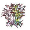
| Title | AMC016 v4.2 in complex with FP-A pAb from animal RQk18 at week 43 | |||||||||
 Map data Map data | ||||||||||
 Sample Sample |
| |||||||||
 Keywords Keywords | HIV-1 / polyclonal / cryoEMPEM / STRUCTURAL PROTEIN / VIRAL PROTEIN-IMMUNE SYSTEM complex | |||||||||
| Biological species |   Human immunodeficiency virus 1 / Human immunodeficiency virus 1 /  | |||||||||
| Method | single particle reconstruction / cryo EM / Resolution: 3.04 Å | |||||||||
 Authors Authors | Pratap PP / Ozorowski G / Ward AB | |||||||||
| Funding support |  United States, 1 items United States, 1 items
| |||||||||
 Citation Citation | Journal: bioRxiv / Year: 2025 Title: Immunofocusing on the conserved fusion peptide of HIV envelope glycoprotein in rhesus macaques. Authors: Payal P Pratap / Christopher A Cottrell / James Quinn / Diane G Carnathan / Daniel L V Bader / Andy S Tran / Chiamaka A Enemuo / Julia T Ngo / Sara T Richey / Hongmei Gao / Xiaoying Shen / ...Authors: Payal P Pratap / Christopher A Cottrell / James Quinn / Diane G Carnathan / Daniel L V Bader / Andy S Tran / Chiamaka A Enemuo / Julia T Ngo / Sara T Richey / Hongmei Gao / Xiaoying Shen / Kelli M Greene / Jonathan Hurtado / Katarzyna Kaczmarek Michaels / Elana Ben-Akiva / Ashley Lemnios / Mariane B Melo / Joel D Allen / Gabriel Ozorowski / Max Crispin / Bryan Briney / David Montefiori / Guido Silvestri / Darrell J Irvine / Shane Crotty / Andrew B Ward /  Abstract: During infection, the fusion peptide (FP) of HIV envelope glycoprotein (Env) serves a central role in viral fusion with the host cell. As such, the FP is highly conserved and therefore an attractive ...During infection, the fusion peptide (FP) of HIV envelope glycoprotein (Env) serves a central role in viral fusion with the host cell. As such, the FP is highly conserved and therefore an attractive epitope for vaccine design. Here, we describe a vaccination study in non-human primates (NHPs) where glycan deletions were made on soluble HIV Env to increase FP epitope exposure. When delivered via implantable osmotic pumps, this immunogen primed immune responses against the FP, which were then boosted with heterologous trimers resulting in a focused immune response targeting the conserved FP epitope. Although autologous immunizations did not elicit high affinity FP-targeting antibodies, the conserved FP epitope on a heterologous trimer further matured the lower affinity, FP-targeting B cells. This study suggests using epitope conservation strategies on distinct Env trimer immunogens can focus humoral responses on desired neutralizing epitopes and suppress immune-distracting antibody responses against non-neutralizing epitopes. | |||||||||
| History |
|
- Structure visualization
| Supplemental images |
|---|

- Downloads & links
-EMDB archive
| Map data | emd_49413.map.gz | 259.2 MB |  EMDB map data format EMDB map data format | |
|---|---|---|---|---|
| Header (meta data) | emd-49413-v30.xmlemd-49413.xml | 24.8 KB 24.8 KB | Display Display | EMDB header |
| FSC (resolution estimation) | emd_49413_fsc.xml | 13.7 KB | Display | FSC data file |
| Images |  emd_49413.png emd_49413.png | 180 KB | ||
| Masks | emd_49413_msk_1.map | 274.6 MB | Mask map | |
| Filedesc metadata | emd-49413.cif.gz | 7.7 KB | ||
| Others | emd_49413_half_map_1.map.gzemd_49413_half_map_2.map.gz | 254.4 MB 254.4 MB | ||
| Archive directory |  http://ftp.pdbj.org/pub/emdb/structures/EMD-49413ftp://ftp.pdbj.org/pub/emdb/structures/EMD-49413 http://ftp.pdbj.org/pub/emdb/structures/EMD-49413ftp://ftp.pdbj.org/pub/emdb/structures/EMD-49413 | HTTPS FTP |
-Related structure data
| Related structure data |  9nhjMC  9nhhC  9nhiC  9nhkC  9nhlC  9nhmC  9nhnC  9nhoC  9ni9C M: atomic model generated by this map C: citing same article ( |
|---|


-Links
| EMDB pages | EMDB (EBI/PDBe) / EMDataResource |
|---|
-Map
| File | Download / File: emd_49413.map.gz / Format: CCP4 / Size: 274.6 MB / Type: IMAGE STORED AS FLOATING POINT NUMBER (4 BYTES) | ||||||||||||||||||||||||||||||||||||
|---|---|---|---|---|---|---|---|---|---|---|---|---|---|---|---|---|---|---|---|---|---|---|---|---|---|---|---|---|---|---|---|---|---|---|---|---|---|
| Projections & slices | Image control
Images are generated by Spider. | ||||||||||||||||||||||||||||||||||||
| Voxel size | X=Y=Z: 1.045 Å | ||||||||||||||||||||||||||||||||||||
| Density |
| ||||||||||||||||||||||||||||||||||||
| Symmetry | Space group: 1 | ||||||||||||||||||||||||||||||||||||
| Details | EMDB XML:
|
 Z (Sec.)
 Y (Row.)
 X (Col.)


-Supplemental data
-Mask #1
| File | emd_49413_msk_1.map | ||||||||||||
|---|---|---|---|---|---|---|---|---|---|---|---|---|---|
| Projections & Slices |
| ||||||||||||
| Density Histograms |


-Half map: #2
| File | emd_49413_half_map_1.map | ||||||||||||
|---|---|---|---|---|---|---|---|---|---|---|---|---|---|
| Projections & Slices |
| ||||||||||||
| Density Histograms |


-Half map: #1
| File | emd_49413_half_map_2.map | ||||||||||||
|---|---|---|---|---|---|---|---|---|---|---|---|---|---|
| Projections & Slices |
| ||||||||||||
| Density Histograms |


- Sample components
-Entire : AMC016 v4.2 in complex with FP-A pAb from animal RQk18 at week 43
| Entire | Name: AMC016 v4.2 in complex with FP-A pAb from animal RQk18 at week 43 |
|---|---|
| Components |
|
-Supramolecule #1: AMC016 v4.2 in complex with FP-A pAb from animal RQk18 at week 43
| Supramolecule | Name: AMC016 v4.2 in complex with FP-A pAb from animal RQk18 at week 43 type: complex / ID: 1 / Parent: 0 / Macromolecule list: #1-#4 |
|---|---|
| Source (natural) | Organism: Human immunodeficiency virus 1 |
| Molecular weight | Theoretical: 0.470 kDa/nm |
-Macromolecule #1: RQk-FP-A pAb heavy chain
| Macromolecule | Name: RQk-FP-A pAb heavy chain / type: protein_or_peptide / ID: 1 / Number of copies: 1 / Enantiomer: LEVO |
|---|---|
| Source (natural) | Organism: |
| Molecular weight | Theoretical: 10.809309 KDa |
| Sequence | String: (UNK)(UNK)(UNK)(UNK)(UNK)(UNK)(UNK)(UNK)(UNK)(UNK) (UNK)(UNK)(UNK)(UNK)(UNK)(UNK) (UNK)(UNK)(UNK) (UNK)(UNK)C(UNK)(UNK)(UNK)(UNK)(UNK)(UNK)(UNK) (UNK)(UNK)(UNK) (UNK)(UNK)(UNK)(UNK)(UNK)(UNK) ...String: (UNK)(UNK)(UNK)(UNK)(UNK)(UNK)(UNK)(UNK)(UNK)(UNK) (UNK)(UNK)(UNK)(UNK)(UNK)(UNK) (UNK)(UNK)(UNK) (UNK)(UNK)C(UNK)(UNK)(UNK)(UNK)(UNK)(UNK)(UNK) (UNK)(UNK)(UNK) (UNK)(UNK)(UNK)(UNK)(UNK)(UNK) (UNK)(UNK)(UNK)(UNK)(UNK)(UNK)(UNK)(UNK)W(UNK) (UNK)(UNK)(UNK)(UNK)(UNK)(UNK)(UNK)(UNK)(UNK) (UNK)(UNK)(UNK)(UNK)(UNK)(UNK)(UNK) (UNK)(UNK) (UNK)(UNK)(UNK)(UNK)(UNK)(UNK)(UNK)(UNK)(UNK)(UNK) (UNK)(UNK)(UNK)(UNK) (UNK)(UNK)(UNK)(UNK)(UNK) (UNK)(UNK)(UNK)(UNK)(UNK)(UNK)(UNK)(UNK)(UNK)(UNK) C (UNK)(UNK)(UNK)(UNK)(UNK)(UNK)(UNK)(UNK) (UNK)(UNK)(UNK)(UNK)(UNK)(UNK)(UNK)(UNK) (UNK) (UNK)(UNK)W(UNK)(UNK)(UNK)(UNK)(UNK)(UNK)(UNK) (UNK) |
-Macromolecule #2: RQk-FP-A pAb light chain
| Macromolecule | Name: RQk-FP-A pAb light chain / type: protein_or_peptide / ID: 2 / Number of copies: 1 / Enantiomer: LEVO |
|---|---|
| Source (natural) | Organism: |
| Molecular weight | Theoretical: 9.153279 KDa |
| Sequence | String: (UNK)(UNK)(UNK)(UNK)(UNK)(UNK)(UNK)(UNK)(UNK)(UNK) (UNK)(UNK)(UNK)(UNK)(UNK)(UNK) (UNK)(UNK)(UNK) (UNK)(UNK)C(UNK)(UNK)(UNK)(UNK)(UNK)(UNK)(UNK) (UNK)(UNK)(UNK) (UNK)W(UNK)(UNK)(UNK)(UNK) ...String: (UNK)(UNK)(UNK)(UNK)(UNK)(UNK)(UNK)(UNK)(UNK)(UNK) (UNK)(UNK)(UNK)(UNK)(UNK)(UNK) (UNK)(UNK)(UNK) (UNK)(UNK)C(UNK)(UNK)(UNK)(UNK)(UNK)(UNK)(UNK) (UNK)(UNK)(UNK) (UNK)W(UNK)(UNK)(UNK)(UNK) (UNK)(UNK)(UNK)(UNK)(UNK)(UNK)(UNK)(UNK)(UNK)(UNK) (UNK)(UNK)(UNK)(UNK)(UNK)(UNK)(UNK)(UNK)(UNK) (UNK)(UNK)(UNK)(UNK)(UNK)(UNK)(UNK) (UNK)(UNK) (UNK)(UNK)(UNK)(UNK)(UNK)(UNK)(UNK)(UNK)(UNK)(UNK) (UNK)(UNK)(UNK)(UNK) (UNK)(UNK)(UNK)(UNK)(UNK) (UNK)C(UNK)(UNK)(UNK)(UNK)(UNK)(UNK)(UNK)(UNK) (UNK)F (UNK)(UNK)(UNK)(UNK)(UNK)(UNK)(UNK) (UNK) |
-Macromolecule #3: AMC016 v4.2 envelope glycoprotein gp120
| Macromolecule | Name: AMC016 v4.2 envelope glycoprotein gp120 / type: protein_or_peptide / ID: 3 / Number of copies: 3 / Enantiomer: LEVO |
|---|---|
| Source (natural) | Organism: Human immunodeficiency virus 1 |
| Molecular weight | Theoretical: 58.713809 KDa |
| Recombinant expression | Organism:  Homo sapiens (human) Homo sapiens (human) |
| Sequence | String: MDAMKRGLCC VLLLCGAVFV SPSQEIHARF RRGARAEEEL WVTVYYGVPV WKEATTTLFC ASDAKAYDTE VHNVWATHCC VPTDPSPQE VVLENVTENF NMWKNNMVEQ MHEDIISLWD QSLKPCVKLT PLCVTLNCTD LGNATDAINR NTTDAPNSTL R TMEEKGEI ...String: MDAMKRGLCC VLLLCGAVFV SPSQEIHARF RRGARAEEEL WVTVYYGVPV WKEATTTLFC ASDAKAYDTE VHNVWATHCC VPTDPSPQE VVLENVTENF NMWKNNMVEQ MHEDIISLWD QSLKPCVKLT PLCVTLNCTD LGNATDAINR NTTDAPNSTL R TMEEKGEI KNCSFNITTS VRDKMQKEYA TFYKLDIVPI DNDNNSYRLI NCNTSVITQA CPKVSFEPIP IHYCAPAGFA IL KCNNKTF NGTGPCTNVS TVQCTHGIRP VVSTQLLLNG SLAEEEIVIR SENFTDNGKT IIVQLNESVE INCTRPNNNT RKS IHIGPG RAFYTTGQII GNIRQAHCNI SRAKWNNTLH KIVKKLREQF RNKTIVFKQS SGGDPEIVMH SFNCGGEFFY CNST QLFNS TWYGNESSDN PGVEGNITLP CRIKQIINLW QEVGKAMYAP PIGGQIRCSS NITGLLLTRD GGNNNITTEI FRPGG GDMR DNWRSELYKY KVVKIEPLGV APTKCKRRVV QRRRRRR |
-Macromolecule #4: AMC016 v4.2 transmembrane protein gp41
| Macromolecule | Name: AMC016 v4.2 transmembrane protein gp41 / type: protein_or_peptide / ID: 4 / Number of copies: 3 / Enantiomer: LEVO |
|---|---|
| Source (natural) | Organism: Human immunodeficiency virus 1 |
| Molecular weight | Theoretical: 17.192521 KDa |
| Recombinant expression | Organism: Homo sapiens (human) |
| Sequence | String: AVGIGAVFLG FLGAAGSTMG AASMTLTVQA RQLLSGIVQQ QSNLLRAPEC QQHLLKDTHW GIKQLQARVL AVEHYLKDQQ LLGIWGCSG KLICTTAVPW NATWSNKTLD NIWNNMTWME WEKEISNYTN LIYNLIEESQ NQQEKNETEN LTLC |
-Macromolecule #8: 2-acetamido-2-deoxy-beta-D-glucopyranose
| Macromolecule | Name: 2-acetamido-2-deoxy-beta-D-glucopyranose / type: ligand / ID: 8 / Number of copies: 44 / Formula: NAG |
|---|---|
| Molecular weight | Theoretical: 221.208 Da |
| Chemical component information |  ChemComp-NAG: |
-Experimental details
-Structure determination
| Method | cryo EM |
|---|---|
 Processing Processing | single particle reconstruction |
| Aggregation state | particle |
-Sample preparation
| Buffer | pH: 7.4 Component:
| ||||||||
|---|---|---|---|---|---|---|---|---|---|
| Grid | Model: Quantifoil / Material: COPPER / Support film - Material: CARBON / Support film - topology: HOLEY / Pretreatment - Type: PLASMA CLEANING | ||||||||
| Vitrification | Cryogen name: ETHANE / Chamber humidity: 100 % / Chamber temperature: 283 K |
- Electron microscopy
| Microscope | TFS KRIOS |
|---|---|
| Image recording | Film or detector model: GATAN K2 SUMMIT (4k x 4k) / Detector mode: COUNTING / Average electron dose: 50.29 e/Å2 |
| Electron beam | Acceleration voltage: 300 kV / Electron source:  FIELD EMISSION GUN FIELD EMISSION GUN |
| Electron optics | Illumination mode: FLOOD BEAM / Imaging mode: BRIGHT FIELD / Nominal defocus max: 2.0 µm / Nominal defocus min: 1.0 µm |
| Sample stage | Specimen holder model: FEI TITAN KRIOS AUTOGRID HOLDER |
| Experimental equipment |  Model: Titan Krios / Image courtesy: FEI Company |