 Movie
Movie Controller

[English] 日本語
 Yorodumi
- EMDB-49351: In situ structure of the cardiac thin filament with troponin from... -
+ Open data

- Basic information
| Entry |  | |||||||||||||||
|---|---|---|---|---|---|---|---|---|---|---|---|---|---|---|---|---|
| Title | In situ structure of the cardiac thin filament with troponin from MYH7(WT/G256E) human induced pluripotent stem cell-derived cardiomyocytes (AICS-0097-141 ACTN2-mEGFP MYH7(WT/G256E)) | |||||||||||||||
 Map data Map data | In situ structure of the cardiac thin filament with troponin from MYH7WT/G256E human induced pluripotent stem cell-derived cardiomyocytes (AICS-0097-141 ACTN2-mEGFP MYH7WT/G256E) | |||||||||||||||
 Sample Sample |
| |||||||||||||||
 Keywords Keywords | cardiac thin filament / CONTRACTILE PROTEIN | |||||||||||||||
| Biological species |  Homo sapiens (human) Homo sapiens (human) | |||||||||||||||
| Method | subtomogram averaging / cryo EM / Resolution: 12.4 Å | |||||||||||||||
 Authors Authors | Woldeyes RA / Chiu W | |||||||||||||||
| Funding support |  United States, 4 items United States, 4 items
| |||||||||||||||
 Citation Citation | Journal: To Be Published Title: Structure of the Thin Filament in Human iPSC-derived Cardiomyocytes and its Response to Heart Disease Authors: Woldeyes RA / Chiu W | |||||||||||||||
| History |
|
- Structure visualization
| Supplemental images |
|---|

- Downloads & links
-EMDB archive
| Map data | emd_49351.map.gz | 23 MB |  EMDB map data format EMDB map data format | |
|---|---|---|---|---|
| Header (meta data) | emd-49351-v30.xmlemd-49351.xml | 15.8 KB 15.8 KB | Display Display | EMDB header |
| FSC (resolution estimation) | emd_49351_fsc.xml | 6.5 KB | Display | FSC data file |
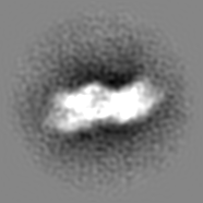
| Images |  emd_49351.png emd_49351.png | 62.9 KB | ||
| Masks | emd_49351_msk_1.map | 24.5 MB | Mask map | |
| Filedesc metadata | emd-49351.cif.gz | 4.2 KB | ||
| Others | emd_49351_half_map_1.map.gzemd_49351_half_map_2.map.gz | 12.5 MB 12.5 MB | ||
| Archive directory |  http://ftp.pdbj.org/pub/emdb/structures/EMD-49351ftp://ftp.pdbj.org/pub/emdb/structures/EMD-49351 http://ftp.pdbj.org/pub/emdb/structures/EMD-49351ftp://ftp.pdbj.org/pub/emdb/structures/EMD-49351 | HTTPS FTP |
-Related structure data


-Links
| EMDB pages | EMDB (EBI/PDBe) / EMDataResource |
|---|
-Map
| File | Download / File: emd_49351.map.gz / Format: CCP4 / Size: 24.5 MB / Type: IMAGE STORED AS FLOATING POINT NUMBER (4 BYTES) | ||||||||||||||||||||||||||||||||||||
|---|---|---|---|---|---|---|---|---|---|---|---|---|---|---|---|---|---|---|---|---|---|---|---|---|---|---|---|---|---|---|---|---|---|---|---|---|---|
| Annotation | In situ structure of the cardiac thin filament with troponin from MYH7WT/G256E human induced pluripotent stem cell-derived cardiomyocytes (AICS-0097-141 ACTN2-mEGFP MYH7WT/G256E) | ||||||||||||||||||||||||||||||||||||
| Projections & slices | Image control
Images are generated by Spider. | ||||||||||||||||||||||||||||||||||||
| Voxel size | X=Y=Z: 2.2 Å | ||||||||||||||||||||||||||||||||||||
| Density |
| ||||||||||||||||||||||||||||||||||||
| Symmetry | Space group: 1 | ||||||||||||||||||||||||||||||||||||
| Details | EMDB XML:
|
 Z (Sec.)
 Y (Row.)
 X (Col.)


-Supplemental data
-Mask #1
| File | emd_49351_msk_1.map | ||||||||||||
|---|---|---|---|---|---|---|---|---|---|---|---|---|---|
| Projections & Slices |
| ||||||||||||
| Density Histograms |


-Half map: #1
| File | emd_49351_half_map_1.map | ||||||||||||
|---|---|---|---|---|---|---|---|---|---|---|---|---|---|
| Projections & Slices |
| ||||||||||||
| Density Histograms |


-Half map: #2
| File | emd_49351_half_map_2.map | ||||||||||||
|---|---|---|---|---|---|---|---|---|---|---|---|---|---|
| Projections & Slices |
| ||||||||||||
| Density Histograms |


- Sample components
-Entire : n situ structure of the cardiac thin filament with troponin from ...
| Entire | Name: n situ structure of the cardiac thin filament with troponin from MYH7(WT/G256E) human induced pluripotent stem cell-derived cardiomyocytes (AICS-0097-141 ACTN2-mEGFP MYH7(WT/G256E)) |
|---|---|
| Components |
|
-Supramolecule #1: n situ structure of the cardiac thin filament with troponin from ...
| Supramolecule | Name: n situ structure of the cardiac thin filament with troponin from MYH7(WT/G256E) human induced pluripotent stem cell-derived cardiomyocytes (AICS-0097-141 ACTN2-mEGFP MYH7(WT/G256E)) type: cell / ID: 1 / Parent: 0 |
|---|---|
| Source (natural) | Organism: Homo sapiens (human) |
-Experimental details
-Structure determination
| Method | cryo EM |
|---|---|
 Processing Processing | subtomogram averaging |
| Aggregation state | cell |
-Sample preparation
| Buffer | pH: 7.4 |
|---|---|
| Vitrification | Cryogen name: ETHANE |
- Electron microscopy
| Microscope | TFS KRIOS |
|---|---|
| Image recording | Film or detector model: GATAN K3 BIOQUANTUM (6k x 4k) / Average electron dose: 2.0 e/Å2 |
| Electron beam | Acceleration voltage: 300 kV / Electron source:  FIELD EMISSION GUN FIELD EMISSION GUN |
| Electron optics | Calibrated magnification: 42000 / Illumination mode: FLOOD BEAM / Imaging mode: BRIGHT FIELD / Cs: 2.7 mm / Nominal defocus max: 5.0 µm / Nominal defocus min: 3.5 µm |
| Sample stage | Specimen holder model: FEI TITAN KRIOS AUTOGRID HOLDER / Cooling holder cryogen: NITROGEN |
| Experimental equipment |  Model: Titan Krios / Image courtesy: FEI Company |

