ジャーナル: Sci Adv / 年: 2020 タイトル: A helical inner scaffold provides a structural basis for centriole cohesion. 著者: Maeva Le Guennec / Nikolai Klena / Davide Gambarotto / Marine H Laporte / Anne-Marie Tassin / Hugo van den Hoek / Philipp S Erdmann / Miroslava Schaffer / Lubomir Kovacik / Susanne Borgers / ...著者: Maeva Le Guennec / Nikolai Klena / Davide Gambarotto / Marine H Laporte / Anne-Marie Tassin / Hugo van den Hoek / Philipp S Erdmann / Miroslava Schaffer / Lubomir Kovacik / Susanne Borgers / Kenneth N Goldie / Henning Stahlberg / Michel Bornens / Juliette Azimzadeh / Benjamin D Engel / Virginie Hamel / Paul Guichard / 要旨: The ninefold radial arrangement of microtubule triplets (MTTs) is the hallmark of the centriole, a conserved organelle crucial for the formation of centrosomes and cilia. Although strong cohesion ...The ninefold radial arrangement of microtubule triplets (MTTs) is the hallmark of the centriole, a conserved organelle crucial for the formation of centrosomes and cilia. Although strong cohesion between MTTs is critical to resist forces applied by ciliary beating and the mitotic spindle, how the centriole maintains its structural integrity is not known. Using cryo-electron tomography and subtomogram averaging of centrioles from four evolutionarily distant species, we found that MTTs are bound together by a helical inner scaffold covering ~70% of the centriole length that maintains MTTs cohesion under compressive forces. Ultrastructure Expansion Microscopy (U-ExM) indicated that POC5, POC1B, FAM161A, and Centrin-2 localize to the scaffold structure along the inner wall of the centriole MTTs. Moreover, we established that these four proteins interact with each other to form a complex that binds microtubules. Together, our results provide a structural and molecular basis for centriole cohesion and geometry.
A: 966.0 Å / B: 966.0 Å / C: 341.55002 Å α=β=γ: 90.0 °
CCP4マップ ヘッダ情報:
mode
Image stored as Reals
Å/pix. X/Y/Z
3.45
3.45
3.45
M x/y/z
280
280
99
origin x/y/z
0.000
0.000
0.000
length x/y/z
966.000
966.000
341.550
α/β/γ
90.000
90.000
90.000
start NX/NY/NZ
0
0
0
NX/NY/NZ
400
400
400
MAP C/R/S
1
2
3
start NC/NR/NS
0
0
0
NC/NR/NS
280
280
99
D min/max/mean
-0.146
2.924
1.146
-
添付データ
-
試料の構成要素
-
全体 : Junction between microtubule triplet of isolated Paramecium tetra...
全体
名称: Junction between microtubule triplet of isolated Paramecium tetraurelia centriole - inner core region
要素
細胞器官・細胞要素: Junction between microtubule triplet of isolated Paramecium tetraurelia centriole - inner core region
-
超分子 #1: Junction between microtubule triplet of isolated Paramecium tetra...
超分子
名称: Junction between microtubule triplet of isolated Paramecium tetraurelia centriole - inner core region タイプ: organelle_or_cellular_component / ID: 1 / 親要素: 0 詳細: This map corresponds to the junction between adjacent microtubule triplets located at the inner core of the centriole
由来(天然)
生物種: Paramecium tetraurelia (ヨツヒメゾウリムシ) Organelle: basal body
-
実験情報
-
構造解析
手法
クライオ電子顕微鏡法
解析
サブトモグラム平均法
試料の集合状態
cell
-
試料調製
緩衝液
pH: 7.2
凍結
凍結剤: ETHANE / 装置: HOMEMADE PLUNGER
-
電子顕微鏡法
顕微鏡
FEI TITAN KRIOS
撮影
フィルム・検出器のモデル: GATAN K2 SUMMIT (4k x 4k) 平均電子線量: 100.0 e/Å2
 ムービー
ムービー コントローラー


 データを開く
 基本情報
基本情報 マップデータ
 試料
試料
 データ登録者
 スイス, 2件
  引用
引用

 構造の表示
構造の表示 ムービービューア
 ダウンロードとリンク
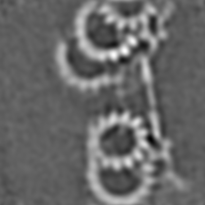 emd_4927.png
 http://ftp.pdbj.org/pub/emdb/structures/EMD-4927


 Z (Sec.)
 Y (Row.)
 X (Col.)


 試料の構成要素
 解析
解析 電子顕微鏡法
 FIELD EMISSION GUN