 Movie
Movie Controller

[English] 日本語
 Yorodumi
- EMDB-49147: Local refinement of crosslinked LetB MCE Rings 5, 6 and 7 (Map 1c) -
+ Open data

- Basic information
| Entry |  | ||||||||||||
|---|---|---|---|---|---|---|---|---|---|---|---|---|---|
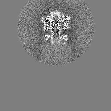
| Title | Local refinement of crosslinked LetB MCE Rings 5, 6 and 7 (Map 1c) | ||||||||||||
 Map data Map data | |||||||||||||
 Sample Sample |
| ||||||||||||
 Keywords Keywords | Lipid transporter / Outer membrane integrity / MCE system / Metal-binding protein / Intermembrane complex / LIPID TRANSPORT | ||||||||||||
| Biological species |  | ||||||||||||
| Method | single particle reconstruction / cryo EM / Resolution: 2.9 Å | ||||||||||||
 Authors Authors | Santarossa CC / Bhabha G / Ekiert DC | ||||||||||||
| Funding support |  United States, 3 items United States, 3 items
| ||||||||||||
 Citation Citation | Journal: bioRxiv / Year: 2025 Title: LetA defines a structurally distinct transporter family involved in lipid trafficking. Authors: Cristina C Santarossa / Yupeng Li / Sara Yousef / Hale S Hasdemir / Carlos C Rodriguez / Max B Haase / Minkyung Baek / Nicolas Coudray / John G Pavek / Kimber N Focke / Annika L Silverberg / ...Authors: Cristina C Santarossa / Yupeng Li / Sara Yousef / Hale S Hasdemir / Carlos C Rodriguez / Max B Haase / Minkyung Baek / Nicolas Coudray / John G Pavek / Kimber N Focke / Annika L Silverberg / Carmelita Bautista / Johannes Yeh / Michael T Marty / David Baker / Emad Tajkhorshid / Damian C Ekiert / Gira Bhabha Abstract: Membrane transport proteins translocate diverse cargos, ranging from small sugars to entire proteins, across cellular membranes. A few structurally distinct protein families have been described that ...Membrane transport proteins translocate diverse cargos, ranging from small sugars to entire proteins, across cellular membranes. A few structurally distinct protein families have been described that account for most of the known membrane transport processes. However, many membrane proteins with predicted transporter functions remain uncharacterized. We determined the structure of LetAB, a phospholipid transporter involved in outer membrane integrity, and found that LetA adopts a distinct architecture that is structurally and evolutionarily unrelated to known transporter families. LetA functions as a pump at one end of a ~225 Å long tunnel formed by its binding partner, MCE protein LetB, creating a pathway for lipid transport between the inner and outer membranes. Unexpectedly, the LetA transmembrane domains adopt a fold that is evolutionarily related to the eukaryotic tetraspanin family of membrane proteins, including TARPs and claudins. LetA has no detectable homology to known transport proteins, and defines a new class of membrane transporters. Through a combination of deep mutational scanning, molecular dynamics simulations, AlphaFold-predicted alternative states, and functional studies, we present a model for how the LetA-like family of membrane transporters may use energy from the proton-motive force to drive the transport of lipids across the bacterial cell envelope. | ||||||||||||
| History |
|
- Structure visualization
| Supplemental images |
|---|

- Downloads & links
-EMDB archive
| Map data | emd_49147.map.gz | 33.1 MB |  EMDB map data format EMDB map data format | |
|---|---|---|---|---|
| Header (meta data) | emd-49147-v30.xmlemd-49147.xml | 19.8 KB 19.8 KB | Display Display | EMDB header |
| Images |  emd_49147.png emd_49147.png | 64.5 KB | ||
| Filedesc metadata | emd-49147.cif.gz | 5.1 KB | ||
| Others | emd_49147_additional_1.map.gzemd_49147_half_map_1.map.gzemd_49147_half_map_2.map.gz | 30.1 MB 59.5 MB 59.5 MB | ||
| Archive directory |  http://ftp.pdbj.org/pub/emdb/structures/EMD-49147ftp://ftp.pdbj.org/pub/emdb/structures/EMD-49147 http://ftp.pdbj.org/pub/emdb/structures/EMD-49147ftp://ftp.pdbj.org/pub/emdb/structures/EMD-49147 | HTTPS FTP |
-Validation report
| Summary document | emd_49147_validation.pdf.gz | 1.2 MB | Display | EMDB validaton report |
|---|---|---|---|---|
| Full document | emd_49147_full_validation.pdf.gz | 1.1 MB | Display | |
| Data in XML | emd_49147_validation.xml.gz | 12.4 KB | Display | |
| Data in CIF | emd_49147_validation.cif.gz | 14.7 KB | Display | |
| Arichive directory | https://ftp.pdbj.org/pub/emdb/validation_reports/EMD-49147ftp://ftp.pdbj.org/pub/emdb/validation_reports/EMD-49147 | HTTPS FTP |
-Related structure data


-Links
| EMDB pages | EMDB (EBI/PDBe) / EMDataResource |
|---|
-Map
| File | Download / File: emd_49147.map.gz / Format: CCP4 / Size: 64 MB / Type: IMAGE STORED AS FLOATING POINT NUMBER (4 BYTES) | ||||||||||||||||||||||||||||||||||||
|---|---|---|---|---|---|---|---|---|---|---|---|---|---|---|---|---|---|---|---|---|---|---|---|---|---|---|---|---|---|---|---|---|---|---|---|---|---|
| Projections & slices | Image control
Images are generated by Spider. | ||||||||||||||||||||||||||||||||||||
| Voxel size | X=Y=Z: 1.02885 Å | ||||||||||||||||||||||||||||||||||||
| Density |
| ||||||||||||||||||||||||||||||||||||
| Symmetry | Space group: 1 | ||||||||||||||||||||||||||||||||||||
| Details | EMDB XML:
|
 Z (Sec.)
 Y (Row.)
 X (Col.)


-Supplemental data
-Additional map: #1
| File | emd_49147_additional_1.map | ||||||||||||
|---|---|---|---|---|---|---|---|---|---|---|---|---|---|
| Projections & Slices |
| ||||||||||||
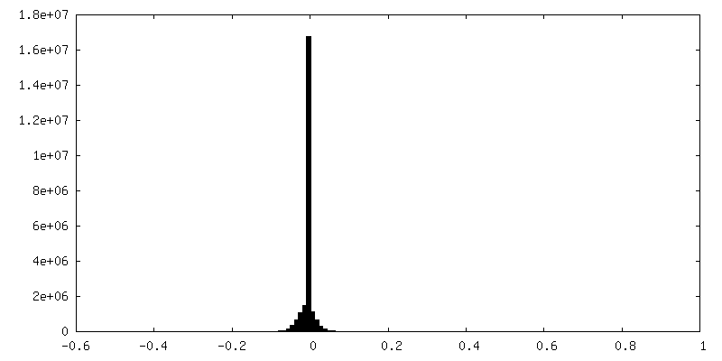
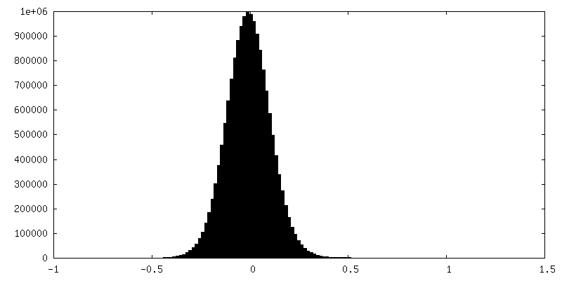
| Density Histograms |


-Half map: #1
| File | emd_49147_half_map_1.map | ||||||||||||
|---|---|---|---|---|---|---|---|---|---|---|---|---|---|
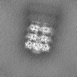
| Projections & Slices |
| ||||||||||||
| Density Histograms |


-Half map: #2
| File | emd_49147_half_map_2.map | ||||||||||||
|---|---|---|---|---|---|---|---|---|---|---|---|---|---|
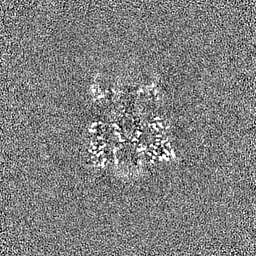
| Projections & Slices |
| ||||||||||||
| Density Histograms |


- Sample components
-Entire : LetB MCE Rings 5, 6 and 7 crosslinked with glutaraldehyde
| Entire | Name: LetB MCE Rings 5, 6 and 7 crosslinked with glutaraldehyde |
|---|---|
| Components |
|
-Supramolecule #1: LetB MCE Rings 5, 6 and 7 crosslinked with glutaraldehyde
| Supramolecule | Name: LetB MCE Rings 5, 6 and 7 crosslinked with glutaraldehyde type: complex / ID: 1 / Parent: 0 / Macromolecule list: #1-#2 |
|---|---|
| Source (natural) | Organism: |
-Experimental details
-Structure determination
| Method | cryo EM |
|---|---|
 Processing Processing | single particle reconstruction |
| Aggregation state | particle |
-Sample preparation
| Concentration | 1 mg/mL | ||||||||||||
|---|---|---|---|---|---|---|---|---|---|---|---|---|---|
| Buffer | pH: 8 Component:
| ||||||||||||
| Grid | Model: Quantifoil R2/2 / Material: COPPER / Mesh: 300 / Support film - Material: CARBON / Support film - topology: CONTINUOUS / Support film - Film thickness: 2 / Pretreatment - Type: GLOW DISCHARGE / Pretreatment - Time: 5 sec. | ||||||||||||
| Vitrification | Cryogen name: ETHANE / Chamber humidity: 100 % / Chamber temperature: 277.15 K / Instrument: FEI VITROBOT MARK IV |
- Electron microscopy
| Microscope | TFS KRIOS |
|---|---|
| Image recording | Film or detector model: GATAN K3 BIOCONTINUUM (6k x 4k) / Number grids imaged: 1 / Number real images: 12464 / Average electron dose: 50.0 e/Å2 |
| Electron beam | Acceleration voltage: 300 kV / Electron source:  FIELD EMISSION GUN FIELD EMISSION GUN |
| Electron optics | Calibrated defocus max: 4.0 µm / Calibrated defocus min: 0.1 µm / Illumination mode: FLOOD BEAM / Imaging mode: BRIGHT FIELD / Cs: 2.7 mm / Nominal defocus max: 2.1 µm / Nominal defocus min: 0.8 µm / Nominal magnification: 81000 |
| Sample stage | Specimen holder model: FEI TITAN KRIOS AUTOGRID HOLDER / Cooling holder cryogen: NITROGEN |
| Experimental equipment |  Model: Titan Krios / Image courtesy: FEI Company |
+Image processing
-Atomic model buiding 1
| Initial model | PDB ID: Chain - Source name: PDB / Chain - Initial model type: experimental model Details: The initial model consisted of the complete biological assembly for PDB entry 6V0E |
|---|---|
| Details | Model was fit as a rigid-body into the individual map using Chimera. Real space refinement was carried out in PHENIX. Models were then manually inspected and adjusted in Coot. Iterative rounds of model building and refinement were performed. |
| Refinement | Space: REAL / Protocol: FLEXIBLE FIT |

