 Movie
Movie Controller

[English] 日本語
 Yorodumi
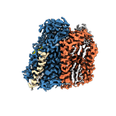
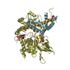
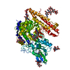
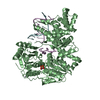
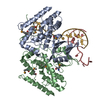
- EMDB-4908: Cryo-EM structure of the E. coli cytochrome bd-I oxidase at 2.68 ... -
+ Open data

- Basic information
| Entry | Database: EMDB / ID: EMD-4908 | |||||||||
|---|---|---|---|---|---|---|---|---|---|---|
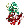
| Title | Cryo-EM structure of the E. coli cytochrome bd-I oxidase at 2.68 A resolution | |||||||||
 Map data Map data | ||||||||||
 Sample Sample |
| |||||||||
 Keywords Keywords | Oxidoreductase Cytochrome bd oxidase bd oxidase Oxidase / MEMBRANE PROTEIN | |||||||||
| Function / homology |  Function and homology information Function and homology informationquinol oxidase (electrogenic, proton-motive force generating) / oxidoreductase activity, acting on diphenols and related substances as donors / cytochrome complex / aerobic electron transport chain / outer membrane / oxidoreductase activity, acting on diphenols and related substances as donors, oxygen as acceptor / oxidative phosphorylation / cellular response to hypoxia / electron transfer activity / heme binding ...quinol oxidase (electrogenic, proton-motive force generating) / oxidoreductase activity, acting on diphenols and related substances as donors / cytochrome complex / aerobic electron transport chain / outer membrane / oxidoreductase activity, acting on diphenols and related substances as donors, oxygen as acceptor / oxidative phosphorylation / cellular response to hypoxia / electron transfer activity / heme binding / membrane / metal ion binding / plasma membrane Similarity search - Function | |||||||||
| Biological species |  | |||||||||
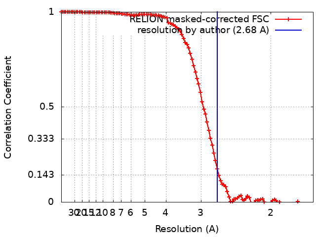
| Method | single particle reconstruction / cryo EM / Resolution: 2.68 Å | |||||||||
 Authors Authors | Safarian S / Hahn A | |||||||||
| Funding support |  Germany, 1 items Germany, 1 items
| |||||||||
 Citation Citation | Journal: Science / Year: 2019 Title: Active site rearrangement and structural divergence in prokaryotic respiratory oxidases. Authors: S Safarian / A Hahn / D J Mills / M Radloff / M L Eisinger / A Nikolaev / J Meier-Credo / F Melin / H Miyoshi / R B Gennis / J Sakamoto / J D Langer / P Hellwig / W Kühlbrandt / H Michel /    Abstract: Cytochrome bd-type quinol oxidases catalyze the reduction of molecular oxygen to water in the respiratory chain of many human-pathogenic bacteria. They are structurally unrelated to mitochondrial ...Cytochrome bd-type quinol oxidases catalyze the reduction of molecular oxygen to water in the respiratory chain of many human-pathogenic bacteria. They are structurally unrelated to mitochondrial cytochrome c oxidases and are therefore a prime target for the development of antimicrobial drugs. We determined the structure of the cytochrome bd-I oxidase by single-particle cryo-electron microscopy to a resolution of 2.7 angstroms. Our structure contains a previously unknown accessory subunit CydH, the L-subfamily-specific Q-loop domain, a structural ubiquinone-8 cofactor, an active-site density interpreted as dioxygen, distinct water-filled proton channels, and an oxygen-conducting pathway. Comparison with another cytochrome bd oxidase reveals structural divergence in the family, including rearrangement of high-spin hemes and conformational adaption of a transmembrane helix to generate a distinct oxygen-binding site. | |||||||||
| History |
|
- Structure visualization
| Movie |
Movie viewer |
|---|---|
| Structure viewer | EM map: SurfViewMolmilJmol/JSmol |
| Supplemental images |
- Downloads & links
-EMDB archive
| Map data | emd_4908.map.gz | 7.9 MB | EMDB map data format | |
|---|---|---|---|---|
| Header (meta data) | emd-4908-v30.xmlemd-4908.xml | 20.5 KB 20.5 KB | Display Display | EMDB header |
| FSC (resolution estimation) | emd_4908_fsc.xml | 10.7 KB | Display | FSC data file |
| Images |  emd_4908.png emd_4908.png | 118.5 KB | ||
| Filedesc metadata | emd-4908.cif.gz | 7 KB | ||
| Archive directory |  http://ftp.pdbj.org/pub/emdb/structures/EMD-4908ftp://ftp.pdbj.org/pub/emdb/structures/EMD-4908 http://ftp.pdbj.org/pub/emdb/structures/EMD-4908ftp://ftp.pdbj.org/pub/emdb/structures/EMD-4908 | HTTPS FTP |
-Related structure data
| Related structure data |  6rkoMC M: atomic model generated by this map C: citing same article ( |
|---|---|
| Similar structure data |


-Links
| EMDB pages | EMDB (EBI/PDBe) / EMDataResource |
|---|
-Map
| File | Download / File: emd_4908.map.gz / Format: CCP4 / Size: 103 MB / Type: IMAGE STORED AS FLOATING POINT NUMBER (4 BYTES) | ||||||||||||||||||||||||||||||||||||||||||||||||||||||||||||
|---|---|---|---|---|---|---|---|---|---|---|---|---|---|---|---|---|---|---|---|---|---|---|---|---|---|---|---|---|---|---|---|---|---|---|---|---|---|---|---|---|---|---|---|---|---|---|---|---|---|---|---|---|---|---|---|---|---|---|---|---|---|
| Projections & slices | Image control
Images are generated by Spider. | ||||||||||||||||||||||||||||||||||||||||||||||||||||||||||||
| Voxel size | X=Y=Z: 0.832 Å | ||||||||||||||||||||||||||||||||||||||||||||||||||||||||||||
| Density |
| ||||||||||||||||||||||||||||||||||||||||||||||||||||||||||||
| Symmetry | Space group: 1 | ||||||||||||||||||||||||||||||||||||||||||||||||||||||||||||
| Details | EMDB XML:
CCP4 map header:
| ||||||||||||||||||||||||||||||||||||||||||||||||||||||||||||
 Z (Sec.)
 Y (Row.)
 X (Col.)


-Supplemental data
- Sample components
+Entire : Cytochrome bd-I oxidase from E. coli
+Supramolecule #1: Cytochrome bd-I oxidase from E. coli
+Macromolecule #1: Cytochrome bd-I ubiquinol oxidase subunit 2
+Macromolecule #2: Cytochrome bd-I ubiquinol oxidase subunit 1
+Macromolecule #3: Uncharacterized protein YnhF
+Macromolecule #4: Cytochrome bd-I ubiquinol oxidase subunit X
+Macromolecule #5: Ubiquinone-8

+Macromolecule #6: (2S)-3-(hexadecanoyloxy)-2-[(9Z)-octadec-9-enoyloxy]propyl 2-(tri...

+Macromolecule #7: CIS-HEME D HYDROXYCHLORIN GAMMA-SPIROLACTONE

+Macromolecule #8: HEME B/C

+Macromolecule #9: OXYGEN MOLECULE
+Macromolecule #10: water

-Experimental details
-Structure determination
| Method | cryo EM |
|---|---|
 Processing Processing | single particle reconstruction |
| Aggregation state | particle |
-Sample preparation
| Concentration | 7 mg/mL | ||||||
|---|---|---|---|---|---|---|---|
| Buffer | pH: 7.5 Component:
| ||||||
| Grid | Model: Quantifoil R1.2/1.3 / Material: COPPER / Mesh: 200 / Pretreatment - Type: GLOW DISCHARGE / Pretreatment - Time: 45 sec. / Pretreatment - Atmosphere: AIR | ||||||
| Vitrification | Cryogen name: ETHANE / Chamber humidity: 100 % / Chamber temperature: 277 K / Instrument: FEI VITROBOT MARK IV |
- Electron microscopy
| Microscope | FEI TITAN KRIOS |
|---|---|
| Temperature | Min: 70.0 K / Max: 70.0 K |
| Image recording | Film or detector model: FEI FALCON III (4k x 4k) / Detector mode: COUNTING / Number grids imaged: 1 / Number real images: 5463 / Average exposure time: 2.0 sec. / Average electron dose: 40.0 e/Å2 |
| Electron beam | Acceleration voltage: 300 kV / Electron source:  FIELD EMISSION GUN FIELD EMISSION GUN |
| Electron optics | C2 aperture diameter: 70.0 µm / Calibrated defocus max: 2.5 µm / Calibrated defocus min: 0.5 µm / Calibrated magnification: 96000 / Illumination mode: FLOOD BEAM / Imaging mode: BRIGHT FIELD / Cs: 2.7 mm / Nominal defocus max: 2.5 µm / Nominal defocus min: 0.5 µm / Nominal magnification: 96000 |
| Sample stage | Specimen holder model: FEI TITAN KRIOS AUTOGRID HOLDER / Cooling holder cryogen: NITROGEN |
| Experimental equipment |  Model: Titan Krios / Image courtesy: FEI Company |
+Image processing
-Atomic model buiding 1
| Refinement | Space: REAL / Protocol: AB INITIO MODEL / Overall B value: 120 / Target criteria: Cross-correlation coefficient |
|---|---|
| Output model | PDB-6rko: |
