 ムービー
ムービー コントローラー


+ データを開く

- 基本情報
基本情報
| 登録情報 | データベース: EMDB / ID: EMD-4870 | |||||||||
|---|---|---|---|---|---|---|---|---|---|---|
| タイトル | In situ cryo-electron tomogram after cryo-FIB lift-out (from lamella 2) | |||||||||
 マップデータ マップデータ | In situ cryo-electron tomogram after cryo-FIB lift-out (from lamella 2) | |||||||||
 試料 試料 |
| |||||||||
| 生物種 |  | |||||||||
| 手法 | 電子線トモグラフィー法 / クライオ電子顕微鏡法 | |||||||||
 データ登録者 データ登録者 | Schaffer M / Pfeffer S / Mahamid J / Albert S / Plitzko JM | |||||||||
| 資金援助 |  ドイツ, 2件 ドイツ, 2件
| |||||||||
 引用 引用 | ジャーナル: Nat Methods / 年: 2019 タイトル: A cryo-FIB lift-out technique enables molecular-resolution cryo-ET within native Caenorhabditis elegans tissue. 著者: Miroslava Schaffer / Stefan Pfeffer / Julia Mahamid / Stephan Kleindiek / Tim Laugks / Sahradha Albert / Benjamin D Engel / Andreas Rummel / Andrew J Smith / Wolfgang Baumeister / Juergen M Plitzko / 要旨: Cryo-focused ion beam milling of frozen-hydrated cells has recently provided unprecedented insights into the inner space of cells. In combination with cryo-electron tomography, this method allows ...Cryo-focused ion beam milling of frozen-hydrated cells has recently provided unprecedented insights into the inner space of cells. In combination with cryo-electron tomography, this method allows access to native structures deep inside cells, enabling structural studies of macromolecules in situ. However, this approach has been mainly limited to individual cells that can be completely vitrified by plunge-freezing. Here, we describe a preparation method that is based on the targeted extraction of material from high-pressure-frozen bulk specimens with a cryo-gripper tool. This lift-out technique enables cryo-electron tomography to be performed on multicellular organisms and tissue, extending the range of applications for in situ structural biology. We demonstrate the potential of the lift-out technique with a structural study of cytosolic 80S ribosomes in a Caenorhabditis elegans worm. The preparation quality allowed for subtomogram analysis with sufficient resolution to distinguish individual ribosomal translocation states and revealed significant cell-to-cell variation in ribosome structure. | |||||||||
| 履歴 |
|
- 構造の表示
構造の表示
| ムービー |
 ムービービューア ムービービューア |
|---|---|
| 添付画像 |

- ダウンロードとリンク
-EMDBアーカイブ
| マップデータ | emd_4870.map.gz | 373.1 MB | EMDBマップデータ形式 | |
|---|---|---|---|---|
| ヘッダ (付随情報) | emd-4870-v30.xmlemd-4870.xml | 9.7 KB 9.7 KB | 表示 表示 | EMDBヘッダ |
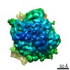
| 画像 | 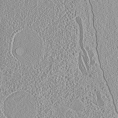 emd_4870.png emd_4870.png | 143.3 KB | ||
| アーカイブディレクトリ |  http://ftp.pdbj.org/pub/emdb/structures/EMD-4870ftp://ftp.pdbj.org/pub/emdb/structures/EMD-4870 http://ftp.pdbj.org/pub/emdb/structures/EMD-4870ftp://ftp.pdbj.org/pub/emdb/structures/EMD-4870 | HTTPS FTP |
-検証レポート
| 文書・要旨 | emd_4870_validation.pdf.gz | 212.9 KB | 表示 | EMDB検証レポート |
|---|---|---|---|---|
| 文書・詳細版 | emd_4870_full_validation.pdf.gz | 212 KB | 表示 | |
| XML形式データ | emd_4870_validation.xml.gz | 4.4 KB | 表示 | |
| アーカイブディレクトリ | https://ftp.pdbj.org/pub/emdb/validation_reports/EMD-4870ftp://ftp.pdbj.org/pub/emdb/validation_reports/EMD-4870 | HTTPS FTP |
-関連構造データ
-リンク
| EMDBのページ | EMDB (EBI/PDBe) / EMDataResource |
|---|
-マップ
| ファイル | ダウンロード / ファイル: emd_4870.map.gz / 形式: CCP4 / 大きさ: 762.2 MB / タイプ: IMAGE STORED AS SIGNED INTEGER (2 BYTES) | ||||||||||||||||||||||||||||||||||||||||||||||||||||||||||||
|---|---|---|---|---|---|---|---|---|---|---|---|---|---|---|---|---|---|---|---|---|---|---|---|---|---|---|---|---|---|---|---|---|---|---|---|---|---|---|---|---|---|---|---|---|---|---|---|---|---|---|---|---|---|---|---|---|---|---|---|---|---|
| 注釈 | In situ cryo-electron tomogram after cryo-FIB lift-out (from lamella 2) | ||||||||||||||||||||||||||||||||||||||||||||||||||||||||||||
| 投影像・断面図 | 画像のコントロール
画像は Spider により作成 これらの図は立方格子座標系で作成されたものです | ||||||||||||||||||||||||||||||||||||||||||||||||||||||||||||
| ボクセルのサイズ | X=Y=Z: 13.68 Å | ||||||||||||||||||||||||||||||||||||||||||||||||||||||||||||
| 密度 |
| ||||||||||||||||||||||||||||||||||||||||||||||||||||||||||||
| 対称性 | 空間群: 1 | ||||||||||||||||||||||||||||||||||||||||||||||||||||||||||||
| 詳細 | EMDB XML:
CCP4マップ ヘッダ情報:
| ||||||||||||||||||||||||||||||||||||||||||||||||||||||||||||
 Z (Sec.)
 Y (Row.)
 X (Col.)


-添付データ
- 試料の構成要素
-全体 : Native cellular environment after cryo-FIB lift-out from C. elega...
| 全体 | 名称: Native cellular environment after cryo-FIB lift-out from C. elegans (from lamella 2) |
|---|---|
| 要素 |
|
-超分子 #1: Native cellular environment after cryo-FIB lift-out from C. elega...
| 超分子 | 名称: Native cellular environment after cryo-FIB lift-out from C. elegans (from lamella 2) タイプ: tissue / ID: 1 / 親要素: 0 |
|---|---|
| 由来(天然) | 生物種: |
-実験情報
-構造解析
| 手法 | クライオ電子顕微鏡法 |
|---|---|
 解析 解析 | 電子線トモグラフィー法 |
| 試料の集合状態 | tissue |
-試料調製
| 緩衝液 | pH: 7.3 |
|---|---|
| 凍結 | 凍結剤: NITROGEN |
| 詳細 | Adult C. elegans worms. |
| 加圧凍結法 | 装置: OTHER 詳細: The value given for _emd_high_pressure_freezing.instrument is BAL-TEC HPM 100 (Leica). This is not in a list of allowed values set(['LEICA EM PACT2', 'LEICA EM PACT', 'EMS-002 RAPID IMMERSION ...詳細: The value given for _emd_high_pressure_freezing.instrument is BAL-TEC HPM 100 (Leica). This is not in a list of allowed values set(['LEICA EM PACT2', 'LEICA EM PACT', 'EMS-002 RAPID IMMERSION FREEZER', 'OTHER', 'LEICA EM HPM100', 'BAL-TEC HPM 010']) so OTHER is written into the XML file. |
| 切片作成 | 集束イオンビーム - 装置: OTHER / 集束イオンビーム - イオン: OTHER / 集束イオンビーム - 電圧: 30 kV / 集束イオンビーム - 電流: 0.05 nA / 集束イオンビーム - 時間: 3500 sec. / 集束イオンビーム - 温度: 92 K / 集束イオンビーム - Initial thickness: 3000 nm / 集束イオンビーム - 最終 厚さ: 150 nm 集束イオンビーム - 詳細: FIB instrument equipped with a Quorum PP3000T cryo-system (Quorum Technologies) and a Kleindiek MM3A-EM micromanipulator (Kleindiek Nanotechnik GmbH).. The value ...集束イオンビーム - 詳細: FIB instrument equipped with a Quorum PP3000T cryo-system (Quorum Technologies) and a Kleindiek MM3A-EM micromanipulator (Kleindiek Nanotechnik GmbH).. The value given for _emd_sectioning_focused_ion_beam.instrument is FIB Quanta 3D FEG (Thermo Fisher). This is not in a list of allowed values set(['DB235', 'OTHER']) so OTHER is written into the XML file. |
- 電子顕微鏡法
| 顕微鏡 | FEI TITAN KRIOS |
|---|---|
| 撮影 | フィルム・検出器のモデル: GATAN K2 SUMMIT (4k x 4k) 検出モード: COUNTING / 平均電子線量: 1.5 e/Å2 |
| 電子線 | 加速電圧: 300 kV / 電子線源:  FIELD EMISSION GUN FIELD EMISSION GUN |
| 電子光学系 | 照射モード: FLOOD BEAM / 撮影モード: BRIGHT FIELD |
| 実験機器 |  モデル: Titan Krios / 画像提供: FEI Company |
-画像解析
| 最終 再構成 | アルゴリズム: BACK PROJECTION / ソフトウェア - 名称: IMOD / 使用した粒子像数: 39 |
|---|
