 Movie
Movie Controller

[English] 日本語
 Yorodumi
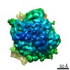
- EMDB-4870: In situ cryo-electron tomogram after cryo-FIB lift-out (from lame... -
+ Open data

- Basic information
| Entry | Database: EMDB / ID: EMD-4870 | |||||||||
|---|---|---|---|---|---|---|---|---|---|---|
| Title | In situ cryo-electron tomogram after cryo-FIB lift-out (from lamella 2) | |||||||||
 Map data Map data | In situ cryo-electron tomogram after cryo-FIB lift-out (from lamella 2) | |||||||||
 Sample Sample |
| |||||||||
| Biological species |  | |||||||||
| Method | electron tomography / cryo EM | |||||||||
 Authors Authors | Schaffer M / Pfeffer S / Mahamid J / Albert S / Plitzko JM | |||||||||
| Funding support |  Germany, 2 items Germany, 2 items
| |||||||||
 Citation Citation | Journal: Nat Methods / Year: 2019 Title: A cryo-FIB lift-out technique enables molecular-resolution cryo-ET within native Caenorhabditis elegans tissue. Authors: Miroslava Schaffer / Stefan Pfeffer / Julia Mahamid / Stephan Kleindiek / Tim Laugks / Sahradha Albert / Benjamin D Engel / Andreas Rummel / Andrew J Smith / Wolfgang Baumeister / Juergen M Plitzko / Abstract: Cryo-focused ion beam milling of frozen-hydrated cells has recently provided unprecedented insights into the inner space of cells. In combination with cryo-electron tomography, this method allows ...Cryo-focused ion beam milling of frozen-hydrated cells has recently provided unprecedented insights into the inner space of cells. In combination with cryo-electron tomography, this method allows access to native structures deep inside cells, enabling structural studies of macromolecules in situ. However, this approach has been mainly limited to individual cells that can be completely vitrified by plunge-freezing. Here, we describe a preparation method that is based on the targeted extraction of material from high-pressure-frozen bulk specimens with a cryo-gripper tool. This lift-out technique enables cryo-electron tomography to be performed on multicellular organisms and tissue, extending the range of applications for in situ structural biology. We demonstrate the potential of the lift-out technique with a structural study of cytosolic 80S ribosomes in a Caenorhabditis elegans worm. The preparation quality allowed for subtomogram analysis with sufficient resolution to distinguish individual ribosomal translocation states and revealed significant cell-to-cell variation in ribosome structure. | |||||||||
| History |
|
- Structure visualization
| Movie |
 Movie viewer Movie viewer |
|---|---|
| Supplemental images |

- Downloads & links
-EMDB archive
| Map data | emd_4870.map.gz | 373.1 MB | EMDB map data format | |
|---|---|---|---|---|
| Header (meta data) | emd-4870-v30.xmlemd-4870.xml | 9.7 KB 9.7 KB | Display Display | EMDB header |
| Images | 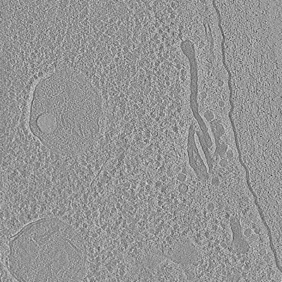 emd_4870.png emd_4870.png | 143.3 KB | ||
| Archive directory |  http://ftp.pdbj.org/pub/emdb/structures/EMD-4870ftp://ftp.pdbj.org/pub/emdb/structures/EMD-4870 http://ftp.pdbj.org/pub/emdb/structures/EMD-4870ftp://ftp.pdbj.org/pub/emdb/structures/EMD-4870 | HTTPS FTP |
-Related structure data
-Links
| EMDB pages | EMDB (EBI/PDBe) / EMDataResource |
|---|
-Map
| File | Download / File: emd_4870.map.gz / Format: CCP4 / Size: 762.2 MB / Type: IMAGE STORED AS SIGNED INTEGER (2 BYTES) | ||||||||||||||||||||||||||||||||||||||||||||||||||||||||||||
|---|---|---|---|---|---|---|---|---|---|---|---|---|---|---|---|---|---|---|---|---|---|---|---|---|---|---|---|---|---|---|---|---|---|---|---|---|---|---|---|---|---|---|---|---|---|---|---|---|---|---|---|---|---|---|---|---|---|---|---|---|---|
| Annotation | In situ cryo-electron tomogram after cryo-FIB lift-out (from lamella 2) | ||||||||||||||||||||||||||||||||||||||||||||||||||||||||||||
| Projections & slices | Image control
Images are generated by Spider. generated in cubic-lattice coordinate | ||||||||||||||||||||||||||||||||||||||||||||||||||||||||||||
| Voxel size | X=Y=Z: 13.68 Å | ||||||||||||||||||||||||||||||||||||||||||||||||||||||||||||
| Density |
| ||||||||||||||||||||||||||||||||||||||||||||||||||||||||||||
| Symmetry | Space group: 1 | ||||||||||||||||||||||||||||||||||||||||||||||||||||||||||||
| Details | EMDB XML:
CCP4 map header:
| ||||||||||||||||||||||||||||||||||||||||||||||||||||||||||||
 Z (Sec.)
 Y (Row.)
 X (Col.)


-Supplemental data
- Sample components
-Entire : Native cellular environment after cryo-FIB lift-out from C. elega...
| Entire | Name: Native cellular environment after cryo-FIB lift-out from C. elegans (from lamella 2) |
|---|---|
| Components |
|
-Supramolecule #1: Native cellular environment after cryo-FIB lift-out from C. elega...
| Supramolecule | Name: Native cellular environment after cryo-FIB lift-out from C. elegans (from lamella 2) type: tissue / ID: 1 / Parent: 0 |
|---|---|
| Source (natural) | Organism: |
-Experimental details
-Structure determination
| Method | cryo EM |
|---|---|
 Processing Processing | electron tomography |
| Aggregation state | tissue |
-Sample preparation
| Buffer | pH: 7.3 |
|---|---|
| Vitrification | Cryogen name: NITROGEN |
| Details | Adult C. elegans worms. |
| High pressure freezing | Instrument: OTHER Details: The value given for _emd_high_pressure_freezing.instrument is BAL-TEC HPM 100 (Leica). This is not in a list of allowed values set(['LEICA EM PACT2', 'LEICA EM PACT', 'EMS-002 RAPID ...Details: The value given for _emd_high_pressure_freezing.instrument is BAL-TEC HPM 100 (Leica). This is not in a list of allowed values set(['LEICA EM PACT2', 'LEICA EM PACT', 'EMS-002 RAPID IMMERSION FREEZER', 'OTHER', 'LEICA EM HPM100', 'BAL-TEC HPM 010']) so OTHER is written into the XML file. |
| Sectioning | Focused ion beam - Instrument: OTHER / Focused ion beam - Ion: OTHER / Focused ion beam - Voltage: 30 kV / Focused ion beam - Current: 0.05 nA / Focused ion beam - Duration: 3500 sec. / Focused ion beam - Temperature: 92 K / Focused ion beam - Initial thickness: 3000 nm / Focused ion beam - Final thickness: 150 nm Focused ion beam - Details: FIB instrument equipped with a Quorum PP3000T cryo-system (Quorum Technologies) and a Kleindiek MM3A-EM micromanipulator (Kleindiek Nanotechnik GmbH).. The value given for ...Focused ion beam - Details: FIB instrument equipped with a Quorum PP3000T cryo-system (Quorum Technologies) and a Kleindiek MM3A-EM micromanipulator (Kleindiek Nanotechnik GmbH).. The value given for _emd_sectioning_focused_ion_beam.instrument is FIB Quanta 3D FEG (Thermo Fisher). This is not in a list of allowed values set(['DB235', 'OTHER']) so OTHER is written into the XML file. |
- Electron microscopy
| Microscope | FEI TITAN KRIOS |
|---|---|
| Image recording | Film or detector model: GATAN K2 SUMMIT (4k x 4k) / Detector mode: COUNTING / Average electron dose: 1.5 e/Å2 |
| Electron beam | Acceleration voltage: 300 kV / Electron source:  FIELD EMISSION GUN FIELD EMISSION GUN |
| Electron optics | Illumination mode: FLOOD BEAM / Imaging mode: BRIGHT FIELD |
| Experimental equipment |  Model: Titan Krios / Image courtesy: FEI Company |
-Image processing
| Final reconstruction | Algorithm: BACK PROJECTION / Software - Name: IMOD / Number images used: 39 |
|---|
