Movie
Movie Controller

[English] 日本語
 Yorodumi
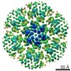
- EMDB-48674: HIV-1 capsid hepta-hexamer templated on small unilamellar vesicles. -
+ Open data

- Basic information
| Entry |  | ||||||||||||
|---|---|---|---|---|---|---|---|---|---|---|---|---|---|
| Title | HIV-1 capsid hepta-hexamer templated on small unilamellar vesicles. | ||||||||||||
 Map data Map data | HIV-1 hepta-hexamer from liposome templating on small unilamellar vesicles. | ||||||||||||
 Sample Sample |
| ||||||||||||
 Keywords Keywords | Capsid / hexamer / HIV-1 / lattice / VIRAL PROTEIN | ||||||||||||
| Biological species |   Human immunodeficiency virus 1 Human immunodeficiency virus 1 | ||||||||||||
| Method | single particle reconstruction / cryo EM / Resolution: 2.98 Å | ||||||||||||
 Authors Authors | Freniere C / Arizaga F / Xiong Y | ||||||||||||
| Funding support |  United States, 3 items United States, 3 items
| ||||||||||||
 Citation Citation | Journal: J Am Chem Soc / Year: 2025 Title: Exploring the Structural Divergence of HIV and SRLV Lentiviral Capsids. Authors: Fidel Arizaga / Christian Freniere / Juan S Rey / Matthew Cook / Chunxiang Wu / Juan R Perilla / Yong Xiong / Abstract: Lentiviruses require a mature capsid to package and traffic their viral genome for successful infection and propagation. Although the HIV-1 capsid structure has been extensively studied, structural ...Lentiviruses require a mature capsid to package and traffic their viral genome for successful infection and propagation. Although the HIV-1 capsid structure has been extensively studied, structural information is lacking for other lentiviral capsids, limiting our understanding. Using cryo-electron microscopy (cryo-EM) and a liposome-templating system, we assembled capsid-like particles (CLPs) and resolved capsid protein (CA) pentamer and hexamer lattice structures from the two major phylogenetic groups of small ruminant lentiviruses (SRLVs). These structures exhibit an overall lattice organization like HIV-1 but differ in key characteristics, notably the absence of inositol hexakisphosphate (IP6) in the SRLV CA lattice─a critical factor for HIV-1 capsid assembly and function. Additionally, SRLV CA pentamers show a unique N-terminal domain orientation, providing insights into SRLV capsid assembly mechanisms. These observations, together with our molecular dynamics (MD) simulation, results suggest a possible mechanism for importing deoxynucleotide triphosphate (dNTP) molecules into SRLV capsids. Furthermore, key regions of host factor interaction, such as the CypA binding motifs, have diverged in the SRLV CA assemblies. Our results contribute to understanding the SRLV lentiviral capsids which may facilitate structure-based inhibitor design strategies. | ||||||||||||
| History |
|
- Structure visualization
| Supplemental images |
|---|

- Downloads & links
-EMDB archive
| Map data | emd_48674.map.gz | 83.1 MB |  EMDB map data format EMDB map data format | |
|---|---|---|---|---|
| Header (meta data) | emd-48674-v30.xmlemd-48674.xml | 17.7 KB 17.7 KB | Display Display | EMDB header |
| FSC (resolution estimation) | emd_48674_fsc.xml | 11.9 KB | Display | FSC data file |
| Images |  emd_48674.png emd_48674.png | 114.3 KB | ||
| Filedesc metadata | emd-48674.cif.gz | 4.8 KB | ||
| Others | emd_48674_half_map_1.map.gzemd_48674_half_map_2.map.gz | 163.6 MB 163.6 MB | ||
| Archive directory |  http://ftp.pdbj.org/pub/emdb/structures/EMD-48674ftp://ftp.pdbj.org/pub/emdb/structures/EMD-48674 http://ftp.pdbj.org/pub/emdb/structures/EMD-48674ftp://ftp.pdbj.org/pub/emdb/structures/EMD-48674 | HTTPS FTP |
-Related structure data


-Links
| EMDB pages | EMDB (EBI/PDBe) / EMDataResource |
|---|
-Map
| File | Download / File: emd_48674.map.gz / Format: CCP4 / Size: 178 MB / Type: IMAGE STORED AS FLOATING POINT NUMBER (4 BYTES) | ||||||||||||||||||||||||||||||||||||
|---|---|---|---|---|---|---|---|---|---|---|---|---|---|---|---|---|---|---|---|---|---|---|---|---|---|---|---|---|---|---|---|---|---|---|---|---|---|
| Annotation | HIV-1 hepta-hexamer from liposome templating on small unilamellar vesicles. | ||||||||||||||||||||||||||||||||||||
| Projections & slices | Image control
Images are generated by Spider. | ||||||||||||||||||||||||||||||||||||
| Voxel size | X=Y=Z: 0.868 Å | ||||||||||||||||||||||||||||||||||||
| Density |
| ||||||||||||||||||||||||||||||||||||
| Symmetry | Space group: 1 | ||||||||||||||||||||||||||||||||||||
| Details | EMDB XML:
|
 Z (Sec.)
 Y (Row.)
 X (Col.)


-Supplemental data
-Half map: half map A
| File | emd_48674_half_map_1.map | ||||||||||||
|---|---|---|---|---|---|---|---|---|---|---|---|---|---|
| Annotation | half map A | ||||||||||||
| Projections & Slices |
| ||||||||||||
| Density Histograms |


-Half map: half map B
| File | emd_48674_half_map_2.map | ||||||||||||
|---|---|---|---|---|---|---|---|---|---|---|---|---|---|
| Annotation | half map B | ||||||||||||
| Projections & Slices |
| ||||||||||||
| Density Histograms |


- Sample components
-Entire : HIV-1 capsid protein p24 with C terminal hexahistidine tag
| Entire | Name: HIV-1 capsid protein p24 with C terminal hexahistidine tag |
|---|---|
| Components |
|
-Supramolecule #1: HIV-1 capsid protein p24 with C terminal hexahistidine tag
| Supramolecule | Name: HIV-1 capsid protein p24 with C terminal hexahistidine tag type: complex / ID: 1 / Parent: 0 Details: Protein was assembled onto small unilamellar vesicles with IP6 at pH 7.4 |
|---|---|
| Source (natural) | Organism: Human immunodeficiency virus 1 |
| Molecular weight | Theoretical: 240 KDa |
-Experimental details
-Structure determination
| Method | cryo EM |
|---|---|
 Processing Processing | single particle reconstruction |
| Aggregation state | 3D array |
-Sample preparation
| Concentration | 4.5 mg/mL | ||||||||||||
|---|---|---|---|---|---|---|---|---|---|---|---|---|---|
| Buffer | pH: 7.4 Component:
Details: Hepes-buffered at pH 7.4 | ||||||||||||
| Grid | Model: Quantifoil R2/1 / Material: COPPER / Mesh: 400 / Support film - Material: CARBON / Support film - topology: HOLEY / Support film - Film thickness: 1 / Pretreatment - Type: GLOW DISCHARGE / Pretreatment - Time: 40 sec. / Details: 11 mA 40s glow discharge in GloCube | ||||||||||||
| Vitrification | Cryogen name: ETHANE / Chamber humidity: 100 % / Chamber temperature: 298 K / Instrument: FEI VITROBOT MARK II / Details: Blot time 6.5 S Blot force 1. | ||||||||||||
| Details | HIV-1 capsid protein templated on small unilamellar vesicles with IP6 |
- Electron microscopy
| Microscope | TFS GLACIOS |
|---|---|
| Temperature | Min: 83.0 K / Max: 103.0 K |
| Image recording | Film or detector model: GATAN K3 (6k x 4k) / Digitization - Dimensions - Width: 6000 pixel / Digitization - Dimensions - Height: 4000 pixel / Number grids imaged: 1 / Number real images: 3800 / Average electron dose: 50.0 e/Å2 |
| Electron beam | Acceleration voltage: 200 kV / Electron source:  FIELD EMISSION GUN FIELD EMISSION GUN |
| Electron optics | C2 aperture diameter: 30.0 µm / Illumination mode: FLOOD BEAM / Imaging mode: BRIGHT FIELD / Cs: 2.7 mm / Nominal defocus max: 20.0 µm / Nominal defocus min: 6.0 µm / Nominal magnification: 45000 |
| Sample stage | Specimen holder model: FEI TITAN KRIOS AUTOGRID HOLDER / Cooling holder cryogen: NITROGEN |