National Institutes of Health/National Institute Of Allergy and Infectious Diseases (NIH/NIAID)
R01 AI127456
United States
Citation
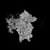
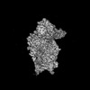
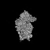
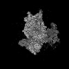
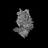
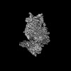
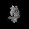
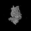
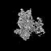
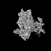
Journal: Nat Microbiol / Year: 2025 Title: Distinct non-canonical translation initiation modes arise for specific host and viral mRNAs during poxvirus-induced shutoff. Authors: Chorong Park / Aaron J Ferrell / Nathan Meade / Peter S Shen / Derek Walsh / Abstract: Many viruses potently inhibit host protein synthesis, termed host shutoff, while employing strategies to sustain their own translation. How and why certain host mRNAs continue to be translated at ...Many viruses potently inhibit host protein synthesis, termed host shutoff, while employing strategies to sustain their own translation. How and why certain host mRNAs continue to be translated at later infection stages remains unclear. Here, using RNAseq and polysome profiling, we show that during shutoff by vaccinia virus (VacV), several host mRNAs increase in polysome occupancy but only a few, primarily JUN that encodes the Jun transcription factor, result in increased protein abundance across multiple cell lines. While dispensable for Jun production, translation of viral mRNAs depended on the small ribosomal protein, Receptor for Activated C Kinase 1 (RACK1) and the eukaryotic Initiation Factor, eIF3. These differential eIF3 dependencies are associated with structurally distinct 5' untranslated regions in viral versus JUN mRNAs. Cryo-electron microscopy structures of 40S ribosomes from mock-infected or VacV-infected cells showed that when bound to eIF3, the rotational range of the RACK1-containing 40S head domain broadens during infection. Our data reveal how eIF3-bound 40S ribosomes are remodelled late in infection and the distinct strategies of translation initiation that arise during shutoff to produce host and viral proteins required for poxvirus spread.
In the structure databanks used in Yorodumi, some data are registered as the other names, "COVID-19 virus" and "2019-nCoV". Here are the details of the virus and the list of structure data.
Jan 31, 2019. EMDB accession codes are about to change! (news from PDBe EMDB page)
EMDB accession codes are about to change! (news from PDBe EMDB page)
The allocation of 4 digits for EMDB accession codes will soon come to an end. Whilst these codes will remain in use, new EMDB accession codes will include an additional digit and will expand incrementally as the available range of codes is exhausted. The current 4-digit format prefixed with “EMD-” (i.e. EMD-XXXX) will advance to a 5-digit format (i.e. EMD-XXXXX), and so on. It is currently estimated that the 4-digit codes will be depleted around Spring 2019, at which point the 5-digit format will come into force.
The EM Navigator/Yorodumi systems omit the EMD- prefix.
Related info.:Q: What is EMD? / ID/Accession-code notation in Yorodumi/EM Navigator
Yorodumi is a browser for structure data from EMDB, PDB, SASBDB, etc.
This page is also the successor to EM Navigator detail page, and also detail information page/front-end page for Omokage search.
The word "yorodu" (or yorozu) is an old Japanese word meaning "ten thousand". "mi" (miru) is to see.
Related info.:EMDB / PDB / SASBDB / Comparison of 3 databanks / Yorodumi Search / Aug 31, 2016. New EM Navigator & Yorodumi / Yorodumi Papers / Jmol/JSmol / Function and homology information / Changes in new EM Navigator and Yorodumi
 Movie
Movie Controller


 Open data
 Basic information
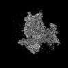
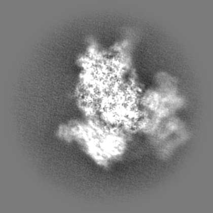
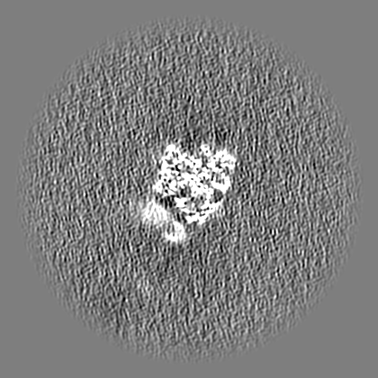
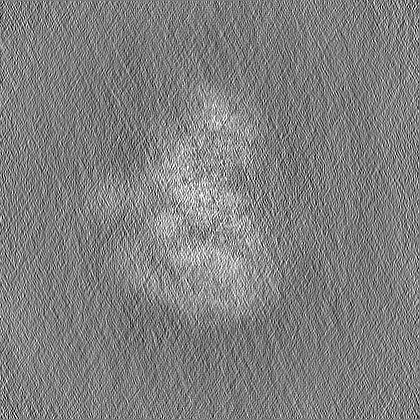
 Map data
 Sample
 Keywords
 Homo sapiens (human)
 Authors
 United States, 1 items
  Citation
 Structure visualization
 Downloads & links
 EMDB map data format
 emd_48642.png
 http://ftp.pdbj.org/pub/emdb/structures/EMD-48642
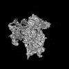
 Z (Sec.)
 Y (Row.)
 X (Col.)


 Sample components
 Processing
 Electron microscopy
 FIELD EMISSION GUN
