 Movie
Movie Controller


+ Open data

- Basic information
| Entry |  | |||||||||
|---|---|---|---|---|---|---|---|---|---|---|
| Title | Cryo-EM structure of KwaA-KwaB complex | |||||||||
 Map data Map data | Cryo-EM density map of KwaAB complex | |||||||||
 Sample Sample |
| |||||||||
 Keywords Keywords | KwaA-KwaB complex / anti-phage defense / IMMUNE SYSTEM | |||||||||
| Function / homology | Protein of unknown function DUF4868 / : / Kiwa protein KwaB-like / : / Anti-phage protein KwaA / defense response to virus / plasma membrane / Kiwa protein KwaA / Kiwa protein KwaB Function and homology information Function and homology information | |||||||||
| Biological species |   Escherichia coli (E. coli) Escherichia coli (E. coli) | |||||||||
| Method | single particle reconstruction / cryo EM / Resolution: 3.75 Å | |||||||||
 Authors Authors | Zhang Z / Patel DJ | |||||||||
| Funding support |  United States, 1 items United States, 1 items
| |||||||||
 Citation Citation | Journal: Cell / Year: 2025 Title: Kiwa is a membrane-embedded defense supercomplex activated at phage attachment sites. Authors: Zhiying Zhang / Thomas C Todeschini / Yi Wu / Roman Kogay / Ameena Naji / Joaquin Cardenas Rodriguez / Rupavidhya Mondi / Daniel Kaganovich / David W Taylor / Jack P K Bravo / Marianna ...Authors: Zhiying Zhang / Thomas C Todeschini / Yi Wu / Roman Kogay / Ameena Naji / Joaquin Cardenas Rodriguez / Rupavidhya Mondi / Daniel Kaganovich / David W Taylor / Jack P K Bravo / Marianna Teplova / Triana Amen / Eugene V Koonin / Dinshaw J Patel / Franklin L Nobrega /  Abstract: Bacteria and archaea deploy diverse antiviral defense systems, many of which remain mechanistically uncharacterized. Here, we characterize Kiwa, a widespread two-component system composed of the ...Bacteria and archaea deploy diverse antiviral defense systems, many of which remain mechanistically uncharacterized. Here, we characterize Kiwa, a widespread two-component system composed of the transmembrane sensor KwaA and the DNA-binding effector KwaB. Cryogenic electron microscopy (cryo-EM) analysis reveals that KwaA and KwaB assemble into a large, membrane-associated supercomplex. Upon phage binding, KwaA senses infection at the membrane, leading to KwaB binding of ejected phage DNA and inhibition of replication and late transcription, without inducing host cell death. Although KwaB can bind DNA independently, its antiviral activity requires association with KwaA, suggesting spatial or conformational regulation. We show that the phage-encoded DNA-mimic protein Gam directly binds and inhibits KwaB but that co-expression with the Gam-targeted RecBCD system restores protection by Kiwa. Our findings support a model in which Kiwa coordinates membrane-associated detection of phage infection with downstream DNA binding by its effector, forming a spatially coordinated antiviral mechanism. | |||||||||
| History |
|
- Structure visualization
| Supplemental images |
|---|

- Downloads & links
-EMDB archive
| Map data | emd_48564.map.gz | 398.1 MB | EMDB map data format | |
|---|---|---|---|---|
| Header (meta data) | emd-48564-v30.xmlemd-48564.xml | 19.1 KB 19.1 KB | Display Display | EMDB header |
| Images |  emd_48564.png emd_48564.png | 23.6 KB | ||
| Filedesc metadata | emd-48564.cif.gz | 5.9 KB | ||
| Others | emd_48564_half_map_1.map.gzemd_48564_half_map_2.map.gz | 390.7 MB 390.7 MB | ||
| Archive directory |  https://data.pdbj.org/pub/emdb/structures/EMD-48564ftp://data.pdbj.org/pub/emdb/structures/EMD-48564 https://data.pdbj.org/pub/emdb/structures/EMD-48564ftp://data.pdbj.org/pub/emdb/structures/EMD-48564 | HTTPS FTP |
-Related structure data
| Related structure data |  9mrxMC  9mrgC  9mrrC  9mtnC  9nyuC  9o0iC M: atomic model generated by this map C: citing same article ( |
|---|---|
| Similar structure data |


-Links
| EMDB pages | EMDB (EBI/PDBe) / EMDataResource |
|---|
-Map
| File | Download / File: emd_48564.map.gz / Format: CCP4 / Size: 421.9 MB / Type: IMAGE STORED AS FLOATING POINT NUMBER (4 BYTES) | ||||||||||||||||||||||||||||||||||||
|---|---|---|---|---|---|---|---|---|---|---|---|---|---|---|---|---|---|---|---|---|---|---|---|---|---|---|---|---|---|---|---|---|---|---|---|---|---|
| Annotation | Cryo-EM density map of KwaAB complex | ||||||||||||||||||||||||||||||||||||
| Projections & slices | Image control
Images are generated by Spider. | ||||||||||||||||||||||||||||||||||||
| Voxel size | X=Y=Z: 0.826 Å | ||||||||||||||||||||||||||||||||||||
| Density |
| ||||||||||||||||||||||||||||||||||||
| Symmetry | Space group: 1 | ||||||||||||||||||||||||||||||||||||
| Details | EMDB XML:
|
 Z (Sec.)
 Y (Row.)
 X (Col.)


-Supplemental data
-Half map: Cryo-EM half A map of KwaAB complex
| File | emd_48564_half_map_1.map | ||||||||||||
|---|---|---|---|---|---|---|---|---|---|---|---|---|---|
| Annotation | Cryo-EM half A map of KwaAB complex | ||||||||||||
| Projections & Slices |
| ||||||||||||
| Density Histograms |


-Half map: Cryo-EM half B map of KwaAB complex
| File | emd_48564_half_map_2.map | ||||||||||||
|---|---|---|---|---|---|---|---|---|---|---|---|---|---|
| Annotation | Cryo-EM half B map of KwaAB complex | ||||||||||||
| Projections & Slices |
| ||||||||||||
| Density Histograms |


- Sample components
-Entire : KwaA-KwaB COMPLEX
| Entire | Name: KwaA-KwaB COMPLEX |
|---|---|
| Components |
|
-Supramolecule #1: KwaA-KwaB COMPLEX
| Supramolecule | Name: KwaA-KwaB COMPLEX / type: complex / ID: 1 / Parent: 0 / Macromolecule list: all |
|---|---|
| Source (natural) | Organism: Escherichia coli (E. coli) |
-Macromolecule #1: Kiwa protein KwaA
| Macromolecule | Name: Kiwa protein KwaA / type: protein_or_peptide / ID: 1 / Number of copies: 4 / Enantiomer: LEVO |
|---|---|
| Source (natural) | Organism: Escherichia coli (E. coli) |
| Molecular weight | Theoretical: 22.52659 KDa |
| Recombinant expression | Organism: Escherichia coli (E. coli) |
| Sequence | String: MQRNTYNKVG LYILSLAMLF VFIIILTAKI PFCFTSDCSF IGLKKLVLTN IVPIVCFVFF LFSIYFYNRL KNITKYNGQD SVKITSCQS ESYESLTFLA TYIVPFMGFS FEDMQKNIAY LLLVVVIGII FIKTDKYYAN PTLALFGFKL YRVNILHPGS G ETKNLIAI ...String: MQRNTYNKVG LYILSLAMLF VFIIILTAKI PFCFTSDCSF IGLKKLVLTN IVPIVCFVFF LFSIYFYNRL KNITKYNGQD SVKITSCQS ESYESLTFLA TYIVPFMGFS FEDMQKNIAY LLLVVVIGII FIKTDKYYAN PTLALFGFKL YRVNILHPGS G ETKNLIAI SNDVLKVDDN VYYSFFDEFV FIARKKI UniProtKB: Kiwa protein KwaA |
-Macromolecule #2: Kiwa protein KwaB
| Macromolecule | Name: Kiwa protein KwaB / type: protein_or_peptide / ID: 2 / Number of copies: 7 / Enantiomer: LEVO |
|---|---|
| Source (natural) | Organism: Escherichia coli (E. coli) |
| Molecular weight | Theoretical: 36.147453 KDa |
| Recombinant expression | Organism: Escherichia coli (E. coli) |
| Sequence | String: MTTQQLKEKI SKIIDNFSGI RVVFTTTANE LKLSRIEGSA LNSIAEGFID KIKEDIINNE DLTSPLLSNF DDRKNALFKF DYEQYPEEF NKITQAIAIP PNSQDYYNPL NKFTDVKGII ILISGDNKCL ALYKNKTNLA VLRNSRKMFN LVPDPDGYLK Q LPNEILRL ...String: MTTQQLKEKI SKIIDNFSGI RVVFTTTANE LKLSRIEGSA LNSIAEGFID KIKEDIINNE DLTSPLLSNF DDRKNALFKF DYEQYPEEF NKITQAIAIP PNSQDYYNPL NKFTDVKGII ILISGDNKCL ALYKNKTNLA VLRNSRKMFN LVPDPDGYLK Q LPNEILRL DFNYDLFSIG EDFYIKNHKT LETQMKFHQV IEAQAVIALN SLRDSLLIED ISGLEKSSRE ISFARKLAKI SK HSPVLGK IDTKTIIDYV SQHKYLSAIL QINEAGDKLL IKTKTSQKHF IKLMSDDYLQ SDLTKIIYMS IAKDRLDE UniProtKB: Kiwa protein KwaB |
-Experimental details
-Structure determination
| Method | cryo EM |
|---|---|
 Processing Processing | single particle reconstruction |
| Aggregation state | particle |
-Sample preparation
| Buffer | pH: 7.5 |
|---|---|
| Vitrification | Cryogen name: ETHANE |
- Electron microscopy
| Microscope | FEI TALOS ARCTICA |
|---|---|
| Image recording | Film or detector model: GATAN K3 (6k x 4k) / Average electron dose: 58.73 e/Å2 |
| Electron beam | Acceleration voltage: 300 kV / Electron source:  FIELD EMISSION GUN FIELD EMISSION GUN |
| Electron optics | Illumination mode: OTHER / Imaging mode: DIFFRACTION / Nominal defocus max: 2.2 µm / Nominal defocus min: 0.8 µm |
| Experimental equipment | 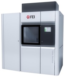 Model: Talos Arctica / Image courtesy: FEI Company |
