 Movie
Movie Controller

[English] 日本語
 Yorodumi
- EMDB-48293: Structure of axonemal DMT from Tetrahymena thermophila RIB22KO mu... -
+ Open data

- Basic information
| Entry |  | |||||||||
|---|---|---|---|---|---|---|---|---|---|---|
| Title | Structure of axonemal DMT from Tetrahymena thermophila RIB22KO mutant, centered on the A-tubule RIB72A/B in 16-nm repeat | |||||||||
 Map data Map data | Structure of axonemal DMT from Tetrahymena thermophila RIB22KO mutant, centered on the A-tubule RIB22 in 16-nm repeat | |||||||||
 Sample Sample |
| |||||||||
 Keywords Keywords | cilia / flagellum / STRUCTURAL PROTEIN | |||||||||
| Biological species |   Tetrahymena thermophila (eukaryote) Tetrahymena thermophila (eukaryote) | |||||||||
| Method | subtomogram averaging / cryo EM / Resolution: 9.64 Å | |||||||||
 Authors Authors | Li S / Winey M | |||||||||
| Funding support |  United States, 1 items United States, 1 items
| |||||||||
 Citation Citation | Journal: Mol Biol Cell / Year: 2025 Title: A ternary complex of MIPs in the A-tubule of basal bodies and axonemes depends on RIB22 and the EF-hand domain of RIB72A in cilia. Authors: Rachel A Howard-Till / Sam Li / Usha Pallabi Kar / Christopher N Fuentes / Amy S Fabritius / Mark Winey / Abstract: The lumens of the highly stable microtubules that make up the core of basal bodies, cilia, and flagella are coated with a network of proteins known as MIPs, or microtubule inner proteins. MIPs are ...The lumens of the highly stable microtubules that make up the core of basal bodies, cilia, and flagella are coated with a network of proteins known as MIPs, or microtubule inner proteins. MIPs are hypothesized to enhance the rigidity and stability of these microtubules, but how they assemble and contribute to cilia function is poorly understood. Here we describe a ciliate specific MIP, RIB22, in . RIB22 is a calmodulin-like protein found in the A-tubule of doublet and triplet microtubules in cilia and basal bodies. Its localization is dependent on the conserved MIP RIB72. Here we use cryogenic electron tomography (cryoET) to examine RIB22 and its interacting partners in axonemes and basal bodies. RIB22 forms a ternary complex with the C-terminal EF-hand domain of RIB72A and another MIP, FAM166A. strains lacking RIB22 or the EF-hand domain of RIB72A showed impaired cilia function. CryoET on axonemes from these strains demonstrated an interdependence of the three proteins for stabilization within the structure. Deletion of the RIB72A EF-hand domain resulted in the apparent loss of multiple MIPs in the region. These findings emphasize the intricacy of the MIP network and the importance of understanding MIPs' functions during cilium assembly and regulation. | |||||||||
| History |
|
- Structure visualization
| Supplemental images |
|---|

- Downloads & links
-EMDB archive
| Map data | emd_48293.map.gz | 14.7 MB |  EMDB map data format EMDB map data format | |
|---|---|---|---|---|
| Header (meta data) | emd-48293-v30.xmlemd-48293.xml | 16.6 KB 16.6 KB | Display Display | EMDB header |
| FSC (resolution estimation) | emd_48293_fsc.xml | 5.8 KB | Display | FSC data file |
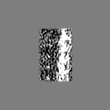
| Images |  emd_48293.png emd_48293.png | 206.5 KB | ||
| Masks | emd_48293_msk_1.map | 15.6 MB | Mask map | |
| Filedesc metadata | emd-48293.cif.gz | 4.7 KB | ||
| Others | emd_48293_half_map_1.map.gzemd_48293_half_map_2.map.gz | 7.7 MB 7.7 MB | ||
| Archive directory |  http://ftp.pdbj.org/pub/emdb/structures/EMD-48293ftp://ftp.pdbj.org/pub/emdb/structures/EMD-48293 http://ftp.pdbj.org/pub/emdb/structures/EMD-48293ftp://ftp.pdbj.org/pub/emdb/structures/EMD-48293 | HTTPS FTP |
-Related structure data


-Links
| EMDB pages | EMDB (EBI/PDBe) / EMDataResource |
|---|
-Map
| File | Download / File: emd_48293.map.gz / Format: CCP4 / Size: 15.6 MB / Type: IMAGE STORED AS FLOATING POINT NUMBER (4 BYTES) | ||||||||||||||||||||||||||||||||||||
|---|---|---|---|---|---|---|---|---|---|---|---|---|---|---|---|---|---|---|---|---|---|---|---|---|---|---|---|---|---|---|---|---|---|---|---|---|---|
| Annotation | Structure of axonemal DMT from Tetrahymena thermophila RIB22KO mutant, centered on the A-tubule RIB22 in 16-nm repeat | ||||||||||||||||||||||||||||||||||||
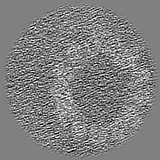
| Projections & slices | Image control
Images are generated by Spider. | ||||||||||||||||||||||||||||||||||||
| Voxel size | X=Y=Z: 2.65 Å | ||||||||||||||||||||||||||||||||||||
| Density |
| ||||||||||||||||||||||||||||||||||||
| Symmetry | Space group: 1 | ||||||||||||||||||||||||||||||||||||
| Details | EMDB XML:
|
 Z (Sec.)
 Y (Row.)
 X (Col.)


-Supplemental data
-Mask #1
| File | emd_48293_msk_1.map | ||||||||||||
|---|---|---|---|---|---|---|---|---|---|---|---|---|---|
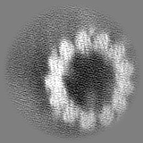
| Projections & Slices |
| ||||||||||||
| Density Histograms |


-Half map: Half map 1
| File | emd_48293_half_map_1.map | ||||||||||||
|---|---|---|---|---|---|---|---|---|---|---|---|---|---|
| Annotation | Half map 1 | ||||||||||||
| Projections & Slices |
| ||||||||||||
| Density Histograms |


-Half map: Half map 2
| File | emd_48293_half_map_2.map | ||||||||||||
|---|---|---|---|---|---|---|---|---|---|---|---|---|---|
| Annotation | Half map 2 | ||||||||||||
| Projections & Slices |
| ||||||||||||
| Density Histograms |


- Sample components
-Entire : Axoneme isolated from Tetrahymena thermophila mutant RIB22KO
| Entire | Name: Axoneme isolated from Tetrahymena thermophila mutant RIB22KO |
|---|---|
| Components |
|
-Supramolecule #1: Axoneme isolated from Tetrahymena thermophila mutant RIB22KO
| Supramolecule | Name: Axoneme isolated from Tetrahymena thermophila mutant RIB22KO type: organelle_or_cellular_component / ID: 1 / Parent: 0 |
|---|---|
| Source (natural) | Organism: Tetrahymena thermophila (eukaryote) |
-Experimental details
-Structure determination
| Method | cryo EM |
|---|---|
 Processing Processing | subtomogram averaging |
| Aggregation state | particle |
-Sample preparation
| Buffer | pH: 7.5 / Details: 1xTE |
|---|---|
| Grid | Model: Quantifoil R2/2 / Material: COPPER/RHODIUM / Mesh: 200 |
| Vitrification | Cryogen name: ETHANE / Instrument: FEI VITROBOT MARK III |
| Details | Axonemes are isolated from a Tetrahymena thermophila mutant RIB22KO |
- Electron microscopy
| Microscope | TFS KRIOS |
|---|---|
| Temperature | Min: 78.0 K / Max: 78.0 K |
| Specialist optics | Energy filter - Name: GIF Bioquantum / Energy filter - Slit width: 20 eV |
| Image recording | Film or detector model: GATAN K3 (6k x 4k) / Digitization - Dimensions - Width: 6000 pixel / Digitization - Dimensions - Height: 4000 pixel / Number real images: 1 / Average exposure time: 0.36 sec. / Average electron dose: 1.33 e/Å2 |
| Electron beam | Acceleration voltage: 300 kV / Electron source:  FIELD EMISSION GUN FIELD EMISSION GUN |
| Electron optics | Calibrated magnification: 18868 / Illumination mode: FLOOD BEAM / Imaging mode: BRIGHT FIELD / Nominal defocus max: 4.0 µm / Nominal defocus min: 2.0 µm / Nominal magnification: 33000 |
| Sample stage | Specimen holder model: FEI TITAN KRIOS AUTOGRID HOLDER / Cooling holder cryogen: NITROGEN |
| Experimental equipment |  Model: Titan Krios / Image courtesy: FEI Company |

