 Movie
Movie Controller

[English] 日本語
 Yorodumi
- EMDB-48260: PI3KC3-C1 bound to RAB1A. Local refinement of VPS34KD in the inac... -
+ Open data

- Basic information
| Entry |  | |||||||||
|---|---|---|---|---|---|---|---|---|---|---|
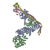
| Title | PI3KC3-C1 bound to RAB1A. Local refinement of VPS34KD in the inactive state, particle subset | |||||||||
 Map data Map data | ||||||||||
 Sample Sample |
| |||||||||
 Keywords Keywords | Complex / GTPase / GTP-binding / Autophagy / Kinase / Lipid Kinase / SIGNALING PROTEIN | |||||||||
| Biological species |  Homo sapiens (human) Homo sapiens (human) | |||||||||
| Method | single particle reconstruction / cryo EM / Resolution: 3.3 Å | |||||||||
 Authors Authors | Cook ASI / Chen M / Hurley JH | |||||||||
| Funding support |  United States, 1 items United States, 1 items
| |||||||||
 Citation Citation | Journal: To Be Published Title: Structural pathway for class III PI 3-kinase activation by the myristoylated GTPbinding pseudokinase VPS15 Authors: Cook ASI / Chen M / Ngyuen TN / Claveras-Cabezudo A / Khuu G / Rao S / Garcia SN / Yang M / Iavarone AT / Ren X / Lazarou M / Hummer G / Hurley JH | |||||||||
| History |
|
- Structure visualization
- Downloads & links
-EMDB archive
| Map data | emd_48260.map.gz | 230.1 MB |  EMDB map data format EMDB map data format | |
|---|---|---|---|---|
| Header (meta data) | emd-48260-v30.xmlemd-48260.xml | 17.8 KB 17.8 KB | Display Display | EMDB header |
| FSC (resolution estimation) | emd_48260_fsc.xml | 13.2 KB | Display | FSC data file |
| Images |  emd_48260.png emd_48260.png | 73.5 KB | ||
| Masks | emd_48260_msk_1.map | 244.1 MB | Mask map | |
| Filedesc metadata | emd-48260.cif.gz | 4.6 KB | ||
| Others | emd_48260_additional_1.map.gzemd_48260_half_map_1.map.gzemd_48260_half_map_2.map.gz | 122.9 MB 226.8 MB 226.8 MB | ||
| Archive directory |  http://ftp.pdbj.org/pub/emdb/structures/EMD-48260ftp://ftp.pdbj.org/pub/emdb/structures/EMD-48260 http://ftp.pdbj.org/pub/emdb/structures/EMD-48260ftp://ftp.pdbj.org/pub/emdb/structures/EMD-48260 | HTTPS FTP |
-Validation report
| Summary document | emd_48260_validation.pdf.gz | 1.2 MB | Display | EMDB validaton report |
|---|---|---|---|---|
| Full document | emd_48260_full_validation.pdf.gz | 1.2 MB | Display | |
| Data in XML | emd_48260_validation.xml.gz | 22.4 KB | Display | |
| Data in CIF | emd_48260_validation.cif.gz | 29 KB | Display | |
| Arichive directory | https://ftp.pdbj.org/pub/emdb/validation_reports/EMD-48260ftp://ftp.pdbj.org/pub/emdb/validation_reports/EMD-48260 | HTTPS FTP |
-Related structure data


-Links
| EMDB pages | EMDB (EBI/PDBe) / EMDataResource |
|---|
-Map
| File | Download / File: emd_48260.map.gz / Format: CCP4 / Size: 244.1 MB / Type: IMAGE STORED AS FLOATING POINT NUMBER (4 BYTES) | ||||||||||||||||||||
|---|---|---|---|---|---|---|---|---|---|---|---|---|---|---|---|---|---|---|---|---|---|
| Voxel size | X=Y=Z: 1.048 Å | ||||||||||||||||||||
| Density |
| ||||||||||||||||||||
| Symmetry | Space group: 1 | ||||||||||||||||||||
| Details | EMDB XML:
|
-Supplemental data
- Sample components
-Entire : Phosphatidylinositol-3 kinase class III complex I bound to RAB1A
| Entire | Name: Phosphatidylinositol-3 kinase class III complex I bound to RAB1A |
|---|---|
| Components |
|
-Supramolecule #1: Phosphatidylinositol-3 kinase class III complex I bound to RAB1A
| Supramolecule | Name: Phosphatidylinositol-3 kinase class III complex I bound to RAB1A type: complex / ID: 1 / Parent: 0 |
|---|---|
| Source (natural) | Organism: Homo sapiens (human) |
| Molecular weight | Theoretical: 384.45 KDa |
-Experimental details
-Structure determination
| Method | cryo EM |
|---|---|
 Processing Processing | single particle reconstruction |
| Aggregation state | particle |
-Sample preparation
| Concentration | 0.2 mg/mL | |||||||||||||||
|---|---|---|---|---|---|---|---|---|---|---|---|---|---|---|---|---|
| Buffer | pH: 7.4 Component:
Details: OG supplemented at time of grid preparation 0.05% | |||||||||||||||
| Grid | Model: Quantifoil Active R2/1 / Material: COPPER / Mesh: 300 / Support film - Material: CARBON / Support film - topology: HOLEY | |||||||||||||||
| Vitrification | Cryogen name: ETHANE / Chamber humidity: 100 % / Chamber temperature: 277 K / Instrument: FEI VITROBOT MARK IV | |||||||||||||||
| Details | monodisperse sample |
- Electron microscopy
| Microscope | TFS KRIOS |
|---|---|
| Specialist optics | Energy filter - Slit width: 20 eV |
| Image recording | Film or detector model: GATAN K3 (6k x 4k) / Number real images: 19300 / Average electron dose: 50.0 e/Å2 |
| Electron beam | Acceleration voltage: 300 kV / Electron source:  FIELD EMISSION GUN FIELD EMISSION GUN |
| Electron optics | Illumination mode: FLOOD BEAM / Imaging mode: BRIGHT FIELD / Cs: 2.7 mm / Nominal defocus max: 2.0 µm / Nominal defocus min: 0.8 µm / Nominal magnification: 81000 |
| Sample stage | Specimen holder model: FEI TITAN KRIOS AUTOGRID HOLDER / Cooling holder cryogen: NITROGEN |
| Experimental equipment |  Model: Titan Krios / Image courtesy: FEI Company |
+Image processing
-Atomic model buiding 1
| Initial model | Chain - Source name: AlphaFold / Chain - Initial model type: in silico model |
|---|---|
| Refinement | Space: REAL / Protocol: FLEXIBLE FIT |
