 Movie
Movie Controller


+ Open data

- Basic information
| Entry | 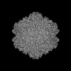 | |||||||||
|---|---|---|---|---|---|---|---|---|---|---|
| Title | Goose Parvovirus Capsid | |||||||||
 Map data Map data | ||||||||||
 Sample Sample |
| |||||||||
 Keywords Keywords | GPV / capsid / cryo-EM / parvovirus / gene therapy / vector / VIRUS LIKE PARTICLE | |||||||||
| Function / homology | Phospholipase A2-like domain / Phospholipase A2-like domain / Parvovirus coat protein VP2 / Parvovirus coat protein VP1/VP2 / Parvovirus coat protein VP2 / Capsid/spike protein, ssDNA virus / T=1 icosahedral viral capsid / structural molecule activity / VP1 Function and homology information Function and homology information | |||||||||
| Biological species |  Goose parvovirus Goose parvovirus | |||||||||
| Method | single particle reconstruction / cryo EM / Resolution: 2.43 Å | |||||||||
 Authors Authors | Jabbari K / Mietzsch M / McKenna R | |||||||||
| Funding support |  United States, 1 items United States, 1 items
| |||||||||
 Citation Citation | Journal: Microorganisms / Year: 2025 Title: The Structural, Biophysical, and Antigenic Characterization of the Goose Parvovirus Capsid. Authors: Korosh Jabbari / Mario Mietzsch / Jane Hsi / Paul Chipman / Jianming Qiu / Robert McKenna / Abstract: Goose parvovirus (GPV) is an etiological agent of Derzsy's disease, afflicting geese and Muscovy ducks worldwide. Its high mortality rate among goslings and ducklings causes large losses to the ...Goose parvovirus (GPV) is an etiological agent of Derzsy's disease, afflicting geese and Muscovy ducks worldwide. Its high mortality rate among goslings and ducklings causes large losses to the waterfowl industry. Toward molecular and structural characterization, virus-like particles (VLPs) of GPV were produced, and the capsid structure was determined by cryogenic electron microscopy (cryo-EM) at a resolution of 2.4 Å. The capsid exhibited structural features conserved among parvoviruses, including surface two-fold depressions, three-fold protrusions, and five-fold channels. A structural comparison of the GPV viral protein (VP) structure with other adeno-associated viruses (AAVs), including human AAV2, AAV5, and quail AAV (QAAV), revealed unique conformations of several surface-accessible variable regions (VRs). Furthermore, the GPV capsid was found to be thermally stable at physiological pH, but less so under lower pH conditions. As a member of the genus , GPV could also be bound by cross-reactive anti-AAV capsid antibodies that bind to the five-fold region of the viruses, as shown by native immuno-dot blot analysis. Finally, the GPV VP structure was compared to those of other bird dependoparvoviruses, which revealed that VR-III may be important for GPV and Muscovy duck parvovirus (MDPV) infection. | |||||||||
| History |
|
- Structure visualization
| Supplemental images |
|---|

- Downloads & links
-EMDB archive
| Map data | emd_48181.map.gz | 442.7 MB | EMDB map data format | |
|---|---|---|---|---|
| Header (meta data) | emd-48181-v30.xmlemd-48181.xml | 13.6 KB 13.6 KB | Display Display | EMDB header |
| Images |  emd_48181.png emd_48181.png | 74.2 KB | ||
| Filedesc metadata | emd-48181.cif.gz | 5.8 KB | ||
| Others | emd_48181_half_map_1.map.gzemd_48181_half_map_2.map.gz | 166.5 MB 166.6 MB | ||
| Archive directory |  http://ftp.pdbj.org/pub/emdb/structures/EMD-48181ftp://ftp.pdbj.org/pub/emdb/structures/EMD-48181 http://ftp.pdbj.org/pub/emdb/structures/EMD-48181ftp://ftp.pdbj.org/pub/emdb/structures/EMD-48181 | HTTPS FTP |
-Validation report
| Summary document | emd_48181_validation.pdf.gz | 1.1 MB | Display | EMDB validaton report |
|---|---|---|---|---|
| Full document | emd_48181_full_validation.pdf.gz | 1.1 MB | Display | |
| Data in XML | emd_48181_validation.xml.gz | 19.6 KB | Display | |
| Data in CIF | emd_48181_validation.cif.gz | 22.3 KB | Display | |
| Arichive directory | https://ftp.pdbj.org/pub/emdb/validation_reports/EMD-48181ftp://ftp.pdbj.org/pub/emdb/validation_reports/EMD-48181 | HTTPS FTP |
-Related structure data
| Related structure data | 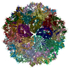 9me0MC M: atomic model generated by this map C: citing same article ( |
|---|---|
| Similar structure data |
-Links
| EMDB pages | EMDB (EBI/PDBe) / EMDataResource |
|---|---|
| Related items in Molecule of the Month |


-Map
| File | Download / File: emd_48181.map.gz / Format: CCP4 / Size: 476.8 MB / Type: IMAGE STORED AS FLOATING POINT NUMBER (4 BYTES) | ||||||||||||||||||||||||||||||||||||
|---|---|---|---|---|---|---|---|---|---|---|---|---|---|---|---|---|---|---|---|---|---|---|---|---|---|---|---|---|---|---|---|---|---|---|---|---|---|
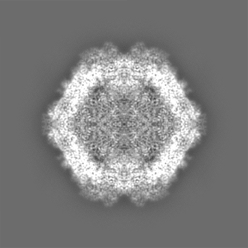
| Projections & slices | Image control
Images are generated by Spider. | ||||||||||||||||||||||||||||||||||||
| Voxel size | X=Y=Z: 0.822 Å | ||||||||||||||||||||||||||||||||||||
| Density |
| ||||||||||||||||||||||||||||||||||||
| Symmetry | Space group: 1 | ||||||||||||||||||||||||||||||||||||
| Details | EMDB XML:
|
 Z (Sec.)
 X (Row.)
 Y (Col.)


-Supplemental data
-Half map: #2
| File | emd_48181_half_map_1.map | ||||||||||||
|---|---|---|---|---|---|---|---|---|---|---|---|---|---|
| Projections & Slices |
| ||||||||||||
| Density Histograms |


-Half map: #1
| File | emd_48181_half_map_2.map | ||||||||||||
|---|---|---|---|---|---|---|---|---|---|---|---|---|---|
| Projections & Slices |
| ||||||||||||
| Density Histograms |


- Sample components
-Entire : Goose parvovirus
| Entire | Name: Goose parvovirus |
|---|---|
| Components |
|
-Supramolecule #1: Goose parvovirus
| Supramolecule | Name: Goose parvovirus / type: virus / ID: 1 / Parent: 0 / Macromolecule list: all / NCBI-ID: 38251 / Sci species name: Goose parvovirus / Virus type: VIRUS-LIKE PARTICLE / Virus isolate: SPECIES / Virus enveloped: No / Virus empty: Yes |
|---|
-Macromolecule #1: VP1
| Macromolecule | Name: VP1 / type: protein_or_peptide / ID: 1 / Number of copies: 60 / Enantiomer: LEVO |
|---|---|
| Source (natural) | Organism: Goose parvovirus |
| Molecular weight | Theoretical: 81.551055 KDa |
| Recombinant expression | Organism:   Spodoptera frugiperda (fall armyworm) Spodoptera frugiperda (fall armyworm) |
| Sequence | String: MSTFLDSFEE WYETAAASWR NLKAGAPQPK PNQQSQSVSP DREPERKDNN RGFVLPGYKY LGPGNGLDKG PPVNKADSVA LEHDKAYDQ QLKAGDNPYI KFNHADQDFI DSLQDDQSFG GNLGKAVFQA KKRILEPFGL VEDPVNTAPA KKNTGKLTDH Y PVVKKPKL ...String: MSTFLDSFEE WYETAAASWR NLKAGAPQPK PNQQSQSVSP DREPERKDNN RGFVLPGYKY LGPGNGLDKG PPVNKADSVA LEHDKAYDQ QLKAGDNPYI KFNHADQDFI DSLQDDQSFG GNLGKAVFQA KKRILEPFGL VEDPVNTAPA KKNTGKLTDH Y PVVKKPKL TEEVSAGGGS SAVQDGGATA EGTEPVAASE MAEGGGGAMG DSSGGADGVG NASGNWHCDS QWMGNTVITK TT RTWVLPS YNNHIYKAIT SGTSQDANVQ YAGYSTPWGY FDFNRFHCHF SPRDWQRLIN NHWGIRPKSL KFKIFNVQVK EVT TQDQTK TIANNLTSTI QVFTDDEHQL PYVLGSATEG TMPPFPSDVY ALPQYGYCTM HTNQNGARFN DRSAFYCLEY FPSQ MLRTG NNFEFTFDFE EVPFHSMFAH SQDLDRLMNP LVDQYLWNFN EVDSSRNAQF KKAVKGAYGT MGRNWLPGPK FLDQR VRAY TGGTDNYANW NIWSNGNKVN LKDRQYLLQP GPVSATYTEG EASSLPAQNI LGIAKDPYRS GSTTAGISDI MVTEEQ EVA PTNGVGWKPY GRTVTNEQNT TTAPTSSDLD VLGALPGMVW QNRDIYLQGP IWAKIPKTDG KFHPSPNLGG FGLHNPP PQ VFIKNTPVPA DPPVEYVHQK WNSYITQYST GQCTVEMVWE LRKENSKRWN PEIQFTSNFS NRTSIMFAPN ETGGYVED R LIGTRYLTQN L UniProtKB: VP1 |
-Experimental details
-Structure determination
| Method | cryo EM |
|---|---|
 Processing Processing | single particle reconstruction |
| Aggregation state | particle |
-Sample preparation
| Buffer | pH: 7.4 |
|---|---|
| Vitrification | Cryogen name: ETHANE |
- Electron microscopy
| Microscope | TFS KRIOS |
|---|---|
| Image recording | Film or detector model: FEI FALCON IV (4k x 4k) / Average electron dose: 50.0 e/Å2 |
| Electron beam | Acceleration voltage: 300 kV / Electron source:  FIELD EMISSION GUN FIELD EMISSION GUN |
| Electron optics | Illumination mode: FLOOD BEAM / Imaging mode: BRIGHT FIELD / Nominal defocus max: 4.7 µm / Nominal defocus min: 0.4 µm |
| Experimental equipment |  Model: Titan Krios / Image courtesy: FEI Company |
-Image processing
| Startup model | Type of model: INSILICO MODEL |
|---|---|
| Final reconstruction | Resolution.type: BY AUTHOR / Resolution: 2.43 Å / Resolution method: FSC 0.143 CUT-OFF / Number images used: 15471 |
| Initial angle assignment | Type: COMMON LINE |
| Final angle assignment | Type: ANGULAR RECONSTITUTION |
