Movie
Movie Controller

[English] 日本語
 Yorodumi
- EMDB-48120: CryoEM structure of H5N1 A/Texas/37/2024 HA bound to Fab 65C6 and... -
+ Open data

- Basic information
| Entry |  | |||||||||
|---|---|---|---|---|---|---|---|---|---|---|
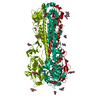
| Title | CryoEM structure of H5N1 A/Texas/37/2024 HA bound to Fab 65C6 and an auto glycan occupying the receptor-binding site | |||||||||
 Map data Map data | ||||||||||
 Sample Sample |
| |||||||||
 Keywords Keywords | H5N1 / antibody / influenza / VIRAL PROTEIN | |||||||||
| Biological species |  Homo sapiens (human) / Homo sapiens (human) /   Influenza A virus Influenza A virus | |||||||||
| Method | single particle reconstruction / cryo EM / Resolution: 2.68 Å | |||||||||
 Authors Authors | Morano NC / Shapiro L / Kwong PD | |||||||||
| Funding support |  United States, 1 items United States, 1 items
| |||||||||
 Citation Citation | Journal: Structure / Year: 2025 Title: Structure of a zoonotic H5N1 hemagglutinin reveals a receptor-binding site occupied by an auto-glycan. Authors: Nicholas C Morano / Yicheng Guo / Jordan E Becker / Zhiteng Li / Jian Yu / David D Ho / Lawrence Shapiro / Peter D Kwong / Abstract: Highly pathogenic avian influenza has spilled into many mammals, most notably cows and poultry, with several dozen human breakthrough infections. Zoonotic crossovers, with hemagglutinins mutated to ...Highly pathogenic avian influenza has spilled into many mammals, most notably cows and poultry, with several dozen human breakthrough infections. Zoonotic crossovers, with hemagglutinins mutated to enhance viral ability to use human α2-6-linked sialic acid receptors versus avian α2-3-linked ones, highlight the pandemic risk. To gain insight into these crossovers, we determined the cryoelectron microscopy (cryo-EM) structure of the hemagglutinin from the zoonotic H5N1 A/Texas/37/2024 strain (clade 2.3.4.4b) in complex with a previously reported neutralizing antibody. Surprisingly, we found that the receptor-binding site of this H5N1 hemagglutinin was already occupied by an α2-3-linked sialic acid and that this glycan emanated from asparagine N169 of a neighboring protomer on hemagglutinin itself. This structure thus highlights recognition by influenza hemagglutinin of an "auto"-α2-3-linked sialic acid from N169, an N-linked glycan conserved in 95% of H5 strains, and adds "auto-glycan recognition," which may play a role in viral dispersal, to the complexities surrounding H5N1 zoonosis. | |||||||||
| History |
|
- Structure visualization
| Supplemental images |
|---|

- Downloads & links
-EMDB archive
| Map data | emd_48120.map.gz | 204 MB |  EMDB map data format EMDB map data format | |
|---|---|---|---|---|
| Header (meta data) | emd-48120-v30.xmlemd-48120.xml | 17.6 KB 17.6 KB | Display Display | EMDB header |
| Images |  emd_48120.png emd_48120.png | 24.7 KB | ||
| Filedesc metadata | emd-48120.cif.gz | 6.6 KB | ||
| Others | emd_48120_half_map_1.map.gzemd_48120_half_map_2.map.gz | 200.4 MB 200.4 MB | ||
| Archive directory |  http://ftp.pdbj.org/pub/emdb/structures/EMD-48120ftp://ftp.pdbj.org/pub/emdb/structures/EMD-48120 http://ftp.pdbj.org/pub/emdb/structures/EMD-48120ftp://ftp.pdbj.org/pub/emdb/structures/EMD-48120 | HTTPS FTP |
-Related structure data

-Links
| EMDB pages | EMDB (EBI/PDBe) / EMDataResource |
|---|
-Map
| File | Download / File: emd_48120.map.gz / Format: CCP4 / Size: 216 MB / Type: IMAGE STORED AS FLOATING POINT NUMBER (4 BYTES) | ||||||||||||||||||||||||||||||||||||
|---|---|---|---|---|---|---|---|---|---|---|---|---|---|---|---|---|---|---|---|---|---|---|---|---|---|---|---|---|---|---|---|---|---|---|---|---|---|
| Projections & slices | Image control
Images are generated by Spider. | ||||||||||||||||||||||||||||||||||||
| Voxel size | X=Y=Z: 0.825 Å | ||||||||||||||||||||||||||||||||||||
| Density |
| ||||||||||||||||||||||||||||||||||||
| Symmetry | Space group: 1 | ||||||||||||||||||||||||||||||||||||
| Details | EMDB XML:
|
 Z (Sec.)
 Y (Row.)
 X (Col.)


-Supplemental data
-Half map: #2
| File | emd_48120_half_map_1.map | ||||||||||||
|---|---|---|---|---|---|---|---|---|---|---|---|---|---|
| Projections & Slices |
| ||||||||||||
| Density Histograms |


-Half map: #1
| File | emd_48120_half_map_2.map | ||||||||||||
|---|---|---|---|---|---|---|---|---|---|---|---|---|---|
| Projections & Slices |
| ||||||||||||
| Density Histograms |


- Sample components
-Entire : Complex of Hemagglutinin from H5N1 HA A/Texas/37/2024 with antibody
| Entire | Name: Complex of Hemagglutinin from H5N1 HA A/Texas/37/2024 with antibody |
|---|---|
| Components |
|
-Supramolecule #1: Complex of Hemagglutinin from H5N1 HA A/Texas/37/2024 with antibody
| Supramolecule | Name: Complex of Hemagglutinin from H5N1 HA A/Texas/37/2024 with antibody type: complex / ID: 1 / Parent: 0 / Macromolecule list: #1-#3 |
|---|---|
| Source (natural) | Organism: Homo sapiens (human) |
| Molecular weight | Theoretical: 360 KDa |
-Macromolecule #1: Hemagglutinin
| Macromolecule | Name: Hemagglutinin / type: protein_or_peptide / ID: 1 / Number of copies: 3 / Enantiomer: LEVO |
|---|---|
| Source (natural) | Organism: Influenza A virus |
| Molecular weight | Theoretical: 65.58575 KDa |
| Recombinant expression | Organism: Homo sapiens (human) |
| Sequence | String: MENIVLLLAI VSLVKSDQIC IGYHANNSTE QVDTIMEKNV TVTHAQDILE KTHNGKLCDL NGVKPLILKD CSVAGWLLGN PMCDEFIRV PEWSYIVERA NPANDLCYPG SLNDYEELKH MLSRINHFEK IQIIPKSSWP NHETSLGVSA ACPYQGAPSF F RNVVWLIK ...String: MENIVLLLAI VSLVKSDQIC IGYHANNSTE QVDTIMEKNV TVTHAQDILE KTHNGKLCDL NGVKPLILKD CSVAGWLLGN PMCDEFIRV PEWSYIVERA NPANDLCYPG SLNDYEELKH MLSRINHFEK IQIIPKSSWP NHETSLGVSA ACPYQGAPSF F RNVVWLIK KNDAYPTIKI SYNNTNREDL LILWGIHHSN NAEEQTNLYK NPITYISVGT STLNQRLAPK IATRSQVNGQ RG RMDFFWT ILKPDDAIHF ESNGNFIAPE YAYKIVKKGD STIMKSGVEY GHCNTKCQTP VGAINSSMPF HNIHPLTIGE CPK YVKSNK LVLATGLRNS PLRRRRRRGL FGAIAGFIEG GWQGMVDGWY GYHHSNEQGS GYAADKESTQ KAIDGVTNKV NSII DKMNT QFEAVGREFN NLERRIENLN KKMEDGFLDV WTYNAELLVL MENERTLDFH DSNVKNLYDK VRLQLRDNAK ELGNG CFEF YHKCDNECME SVRNGTYDYP QYSEEARLKR EGSSGSSGYI PEAPRDGQAY VRKDGEWVLL STFLGHHHHH HHHHGG SGL NDIFEAQKIE WHE |
-Macromolecule #2: Fab 65C6 Heavy Chain
| Macromolecule | Name: Fab 65C6 Heavy Chain / type: protein_or_peptide / ID: 2 / Number of copies: 3 / Enantiomer: LEVO |
|---|---|
| Source (natural) | Organism: Homo sapiens (human) |
| Molecular weight | Theoretical: 25.716766 KDa |
| Recombinant expression | Organism: Homo sapiens (human) |
| Sequence | String: EVQLVQSGAE VKKPGESLRI SCKGFAYSST YFWISWVRQM PGKGLEWMGR IDPTDSYINY SPSFQGHVTI SVDRSISTVY LQWSSLKAS DTAMYYCAYH RRGHFYGSGS AWDWFESWGQ GTLVTVSSAS TKGPSVFPLA PSSKSTSGGT AALGCLVKDY F PEPVTVSW ...String: EVQLVQSGAE VKKPGESLRI SCKGFAYSST YFWISWVRQM PGKGLEWMGR IDPTDSYINY SPSFQGHVTI SVDRSISTVY LQWSSLKAS DTAMYYCAYH RRGHFYGSGS AWDWFESWGQ GTLVTVSSAS TKGPSVFPLA PSSKSTSGGT AALGCLVKDY F PEPVTVSW NSGALTSGVH TFPAVLQSSG LYSLSSVVTV PSSSLGTQTY ICNVNHKPSN TKVDKKVESA SCDKTHTCP |
-Macromolecule #3: Fab 65C6 Light Chain
| Macromolecule | Name: Fab 65C6 Light Chain / type: protein_or_peptide / ID: 3 / Number of copies: 3 / Enantiomer: LEVO |
|---|---|
| Source (natural) | Organism: Homo sapiens (human) |
| Molecular weight | Theoretical: 23.41201 KDa |
| Recombinant expression | Organism: Homo sapiens (human) |
| Sequence | String: EIVLTQSPLT LSVSPGERAT LSCRASQSVS SNLAWYQQMP GQAPRLLIYG ASTRATGIPA RLSGSASGTE FTLTISSLQS EDFAVYYCQ QYNNWPYTFG QGTKLEIKRT VAAPSVFIFP PSDEQLKSGT ASVVCLLNNF YPREAKVQWK VDNALQSGNS Q ESVTEQDS ...String: EIVLTQSPLT LSVSPGERAT LSCRASQSVS SNLAWYQQMP GQAPRLLIYG ASTRATGIPA RLSGSASGTE FTLTISSLQS EDFAVYYCQ QYNNWPYTFG QGTKLEIKRT VAAPSVFIFP PSDEQLKSGT ASVVCLLNNF YPREAKVQWK VDNALQSGNS Q ESVTEQDS KDSTYSLSST LTLSKADYEK HKVYACEVTH QGLSSPVTKS FNRGEC |
-Macromolecule #5: 2-acetamido-2-deoxy-beta-D-glucopyranose
| Macromolecule | Name: 2-acetamido-2-deoxy-beta-D-glucopyranose / type: ligand / ID: 5 / Number of copies: 12 / Formula: NAG |
|---|---|
| Molecular weight | Theoretical: 221.208 Da |
| Chemical component information |  ChemComp-NAG: |
-Experimental details
-Structure determination
| Method | cryo EM |
|---|---|
 Processing Processing | single particle reconstruction |
| Aggregation state | particle |
-Sample preparation
| Buffer | pH: 7.4 |
|---|---|
| Vitrification | Cryogen name: ETHANE |
- Electron microscopy
| Microscope | TFS KRIOS |
|---|---|
| Image recording | Film or detector model: GATAN K3 (6k x 4k) / Average electron dose: 58.0 e/Å2 |
| Electron beam | Acceleration voltage: 300 kV / Electron source:  FIELD EMISSION GUN FIELD EMISSION GUN |
| Electron optics | Illumination mode: FLOOD BEAM / Imaging mode: BRIGHT FIELD / Nominal defocus max: 2.0 µm / Nominal defocus min: 0.8 µm |
| Experimental equipment |  Model: Titan Krios / Image courtesy: FEI Company |
-Image processing
| Startup model | Type of model: PDB ENTRY PDB model - PDB ID: |
|---|---|
| Final reconstruction | Resolution.type: BY AUTHOR / Resolution: 2.68 Å / Resolution method: FSC 0.143 CUT-OFF / Software - Name: cryoSPARC / Number images used: 206541 |
| Initial angle assignment | Type: NOT APPLICABLE |
| Final angle assignment | Type: NOT APPLICABLE |